A dual-energy X-ray absorptiometry (DEXA) scan is a type of low-dose X-ray that indicates the level of bone mineral density (BMD). BMD stands for bone mass density and is a measure of the level of minerals like calcium etc.
A dual-energy X-ray absorptiometry (DEXA) scan is a type of low-dose X-ray that indicates the level of bone mineral density (BMD). BMD stands for bone mass density and is a measure of the level of minerals like calcium etc. in your bones. Low BMD is a risk factor for osteoporosis, a condition that makes bones weak and fragile.
DEXA scans are used to diagnose osteoporosis and to monitor the effectiveness of treatment. They can also be used to assess the risk of fractures in people who are not yet at high risk for osteoporosis.
Why is Dexa Scan Required?
Dexa scans are required for several reasons, primarily related to the evaluation and management of bone health and body composition. Here are some of the key reasons why a Dexa scan may be necessary:
- Diagnosis and monitoring of osteoporosis: Dexa scans are widely used to diagnose osteoporosis and assess an individual's risk of fractures. By measuring bone mineral density (BMD), the scan helps determine if bone density is within the normal range or if there is significant bone loss. The results can guide treatment decisions and monitor the effectiveness of interventions over time.
- Assessment of fracture risk: Dexa scans provide valuable information about an individual's risk of fractures. Low bone density is a significant risk factor for fractures, especially in older adults. The T-score derived from the scan helps identify individuals with a higher risk of fracture and enables healthcare providers to implement preventive measures and interventions.
- Evaluation of body composition: In addition to measuring bone density, Dexa scans can assess body composition by measuring the amount of body fat and lean mass. This information is useful in assessing overall health, identifying the risk of obesity-related complications, and monitoring changes in body composition over time.
- Monitoring the effects of treatments and interventions: Dexa scans are utilized to monitor the effects of interventions aimed at improving bone health, such as medications, lifestyle modifications, and hormone replacement therapies. Regular scans can help track changes in bone density and evaluate the effectiveness of treatments.
- Detection of other conditions: Dexa scans can occasionally reveal other conditions affecting bone health, such as hyperparathyroidism, osteomalacia, and certain types of cancers that may cause bone loss. The scan results can aid in the diagnosis of these conditions and guide further evaluation and management.
It's important to note that the necessity of a Dexa scan depends on individual circumstances and risk factors. Healthcare professionals will determine if a Dexa scan is appropriate based on factors such as age, sex, medical history, and clinical indications.
Uses of DEXA scans
DEXA scans are used for a variety of purposes, including:
- Diagnosing osteoporosis
- Monitoring the effectiveness of osteoporosis treatment
- Assessing the risk of fractures in people who are not yet at high risk for osteoporosis.
- Evaluating the severity of osteoporosis
- Planning surgery for people with osteoporosis
- Measuring the amount of bone loss in people with certain medical conditions, such as rheumatoid arthritis or cancer
- Researching the causes and treatment of osteoporosis
What is a Dexa Scanner?
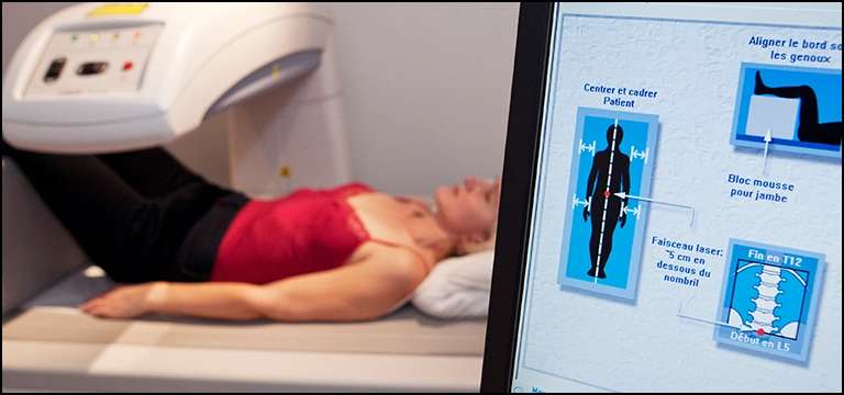
A Dexa scanner, also known as a dual-energy X-ray absorptiometry scanner, is the medical device used to perform Dexa scans. It is a specialized X-ray machine designed to measure bone mineral density (BMD) and body composition.
A Dexa scanner consists of a scanning table, an X-ray tube, and a detector that captures the X-ray images. The patient lies on the scanning table while the scanner passes two X-ray beams through the body. These beams have different energy levels, typically low and high. The scanner measures the amount of X-ray energy that is absorbed by the bones and soft tissues.
The detector records the X-ray images, and the information is sent to a computer system that processes the data and calculates the bone mineral density. The results are typically displayed as a T-score, which compares the patient's BMD to the average BMD of young adults of the same sex.
Modern Dexa scanners are usually compact and have improved features for patient comfort and safety. They emit very low levels of radiation, making the procedure safe and quick. The scanner can be adjusted to focus on specific areas of the body, such as the spine, hip, or forearm, depending on the clinical needs.
Dexa scanners are typically found in radiology departments of hospitals, clinics, and specialized imaging centers. They are operated by trained technicians or radiologists who interpret the results. Dexa scanners play a crucial role in diagnosing and monitoring conditions such as osteoporosis, assessing fracture risk, and evaluating body composition.
Procedure for a DEXA scan
A DEXA scan is comparatively painless as well as quick process. Your body will be scanned while you are lying on a table. Two low-dose X-rays are released by the scanner, and they pass through your bones and soft tissues. Your BMD is determined by the scanner using data on the quantity of X-ray energy absorbed by your bones.
Typically, the entire process takes under ten minutes. Before the scan, you could be requested to take off your shoes, belt, and jewellery. Another requirement could be to put on a hospital gown.
Preparation for a DEXA scan
No special preparation is required for DEXA scan. You do not need to fast or take any medications before the scan.
If you are pregnant, you should tell your doctor before you have a DEXA scan. The amount of radiation exposure from a DEXA scan is very low, but it is always best to be cautious during pregnancy.
Risks of DEXA scans
The risks of DEXA scans are very low. The amount of radiation exposure from a DEXA scan is equivalent to about 1 year of natural background radiation.
In rare cases, DEXA scans can cause allergic reactions to the contrast dye that is sometimes used to improve the image quality.
Results of a DEXA scan
The results of a DEXA scan are reported as a T-score and a Z-score.
- Your BMD is compared to the BMD of young individuals in good health for the T-score. T-scores of -1 and higher are regarded as normal. If the score is between -1 and -2.5, this score indicates low bone mass. Osteoporosis is characterised by a T-score of -2.5 or less.
- The Z-score compares your BMD to the BMD of people of your same age and sex. A Z-score of -1 or higher is considered normal. A Z-score of -1 to -2.5 is considered low bone mass for your age and sex. A Z-score of -2.5 or lower is considered osteoporosis for your age and sex.
Your doctor will discuss the results of your DEXA scan with you and recommend treatment if necessary.
Cost of a DEXA scan
The cost of a DEXA scan varies depending on where you live and the type of insurance you have. In the United States, the average cost of a DEXA scan is about 2000 - 4000 INR.
If you do not have insurance, you may be able to get a DEXA scan at a reduced cost or for free. There are many organizations that offer free or low-cost DEXA scans to people who are at risk for osteoporosis.
Where to get a DEXA scan
DEXA scans are available at many hospitals, clinics, and doctor's offices. You can find a DEXA scan provider near you by using the National Osteoporosis Foundation's (NOF) “Find a DEXA Scan” tool.
The NOF's Find a DEXA Scan tool allows you to search for DEXA scan providers by location, insurance, and other criteria. You can also use the tool to find out if there are any free or low-cost DEXA scan programs available in your area.
Conclusion
In conclusion, a Dexa scan, or dual-energy X-ray absorptiometry scan, is a valuable medical imaging technique used to measure bone mineral density and assess body composition. This non-invasive procedure provides crucial information for the diagnosis, monitoring, and management of conditions such as osteoporosis and related fractures.
By accurately measuring bone density, Dexa scans help healthcare professionals identify individuals at risk of fractures and guide appropriate treatment interventions. The T-score derived from the scan assists in evaluating bone health by comparing it to the average density of young adults. Additionally, Dexa scans offer insights into body composition, including body fat and lean mass measurements, aiding in the assessment of overall health and obesity-related risks.
The Dexa scanner itself is a specialized X-ray machine that emits low levels of radiation and consists of a scanning table, X-ray tube, and detector. With advancements in technology, Dexa scanners have become more compact, comfortable, and efficient, facilitating quick and safe scans.
The use of Dexa scanners is essential in clinical practice, particularly in fields like rheumatology, endocrinology, and geriatrics. It allows healthcare professionals to make informed decisions regarding treatment options, monitor the effects of interventions, and detect other conditions affecting bone health.
Overall, Dexa scans play a crucial role in promoting bone health, preventing fractures, and assessing body composition, contributing to improved patient care and well-being.


