Cervical spine MRI is a non-invasive imaging technique that uses powerful magnets and radio waves to create detailed images of the structures within the neck region. It provides valuable information about the bones, discs,...
Table of contents:
- Introduction
- Understanding Cervical Spine MRI
- The Cervical Spine MRI Procedure
- Preparing for a Cervical Spine MRI
- Potential Findings from Cervical Spine MRI
- Conclusion
- FAQs
Introduction:
The cervical spine, consisting of the seven vertebrae in the neck region, plays a crucial role in providing support, stability, and flexibility to the human body. When individuals experience neck pain, numbness, tingling sensations, or other cervical-related symptoms, healthcare professionals often turn to cervical spine MRI (Magnetic Resonance Imaging) to uncover the underlying causes.
Here, we will explore the various aspects of cervical spine MRI, including its purpose, procedure, preparation, and potential findings.
Whether you are a patient scheduled for a cervical spine MRI or simply curious about this diagnostic tool, we’ll unravel everything surrounding this important medical imaging technique.
Understanding Cervical Spine MRI
Cervical spine MRI is a non-invasive imaging technique that uses powerful magnets and radio waves to create detailed images of the structures within the neck region. It provides valuable information about the bones, discs, nerves, and soft tissues of the cervical spine. By capturing cross-sectional images, MRI can help identify abnormalities, such as herniated discs, spinal cord compression, tumors, infections, or degenerative conditions, which may be causing pain, discomfort, or neurological symptoms in the neck area.
The Cervical Spine MRI Procedure
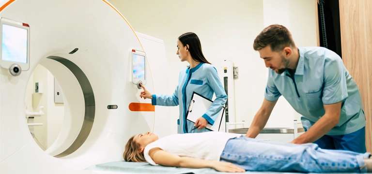
A cervical spine MRI is typically performed in a specialized imaging center or hospital. The patient lies down on a movable examination table, which slides into the MRI machine. To ensure accurate imaging, it is necessary to remain as still as possible during the procedure. The technician may provide earplugs or headphones to minimize the noise generated by the MRI machine.
Before the scan begins, the technician may place a coil or special device around the neck area to enhance the image quality of the cervical spine. The machine emits a strong magnetic field and radio waves, which cause the hydrogen atoms in the body's cells to align. As these atoms return to their original state, they emit signals that are detected by the MRI machine and used to construct detailed images.
The procedure typically takes approximately 30 to 60 minutes, during which multiple images are acquired from different angles and positions. It is important to communicate with the technician through an intercom system, as they will guide you and address any concerns during the scan.
Preparing for a Cervical Spine MRI
Proper preparation ensures a successful and accurate cervical spine MRI.
Here are few general rules to follow:
Communicate with your healthcare provider: Inform your healthcare provider about any pre-existing medical conditions, allergies, or implanted devices, such as pacemakers or metal implants. Some conditions or devices may require special considerations or alternatives to MRI.
Remove metal objects: Before the scan, remove all metal objects, including jewelry, watches, hairpins, and clothing with metallic components, as they can interfere with the MRI's magnetic field.
Discuss medication and sedation: If you experience claustrophobia or anxiety, consult with your healthcare provider about the possibility of medication or sedation to help you relax during the procedure.
Wear comfortable clothing: Opt for loose-fitting, comfortable clothing without metal fasteners or zippers. In some cases, a hospital gown may be provided.
Potential Findings from Cervical Spine MRI
A cervical spine MRI can reveal a host of conditions and abnormalities in a patient.
Some potential findings include:
Herniated Discs: MRI can visualize herniated or bulging discs, which occur when the cushioning discs between the vertebrae protrude or rupture, potentially causing nerve compression and pain.
Spinal Stenosis: This condition refers to the narrowing of the spinal canal, which can compress the spinal cord and nerves. MRI can help identify the location and severity of spinal Stenosis.
Degenerative Disc Disease: MRI can detect signs of degeneration in the spinal discs, such as loss of disc height, disc desiccation (drying out), and the formation of bone spurs.
Tumors and Infections: MRI can identify the presence and location of tumors, abscesses, or infections in the cervical spine.
Spinal Cord Compression: MRI can visualize compression or impingement of the spinal cord or nerve roots, providing critical information for treatment planning.
Cervical spine MRI price
The cost of a cervical spine MRI can vary depending on factors such as geographic location, healthcare facility, and insurance coverage.
Cervical spine MRI price varies in different regions or countries.
Healthcare facilities may offer package deals or discounted rates for multiple imaging studies, including cervical spine MRI.
Insurance coverage can play a major role in determining out-of-pocket expenses for a cervical spine MRI price.
Some healthcare facilities provide financial assistance programs or payment plans to help manage the cost of cervical spine MRI for patients without insurance or with high deductibles.
One should inquire about the price quote and what is included in it. It might be possible that additional charges such as radiologist interpretation or contrast agents could be separate.
Patients shopping around and comparing prices from different healthcare facilities can help patients find the best value for their cervical spine MRI.
Discussing the cost and potential alternatives with your healthcare provider can provide insights into affordability and potential cost-saving measures.
Medical tourism in countries known for affordable healthcare may offer cost-effective options for cervical spine MRI, but it is crucial to consider factors like quality, safety, and travel expenses.
Understanding the cost breakdown and negotiating with healthcare facilities can sometimes result in more favorable pricing for a cervical spine MRI.
Conclusion
A cervical spine MRI is a valuable diagnostic tool that enables healthcare professionals to evaluate and diagnose various cervical spine conditions accurately. It offers detailed images of the structures within the neck region, aiding in the identification of abnormalities, injuries, and diseases.
By understanding the purpose, procedure, and potential findings of cervical spine MRI, patients can feel more informed and confident during the imaging process. If you are experiencing neck pain, or neurological symptoms, or have been advised to undergo a cervical spine MRI, consult with your healthcare provider to determine if this imaging technique is appropriate for your specific situation.
FAQs:
Is cervical spine MRI painful?
No. It is a non-invasive procedure and does not cause any pain. However, some individuals may feel slight discomfort from lying still for an extended period or from the noise generated by the MRI machine. One should let the technician know if there is any discomfort during the scan.
How long does a cervical spine MRI take?
The duration of a cervical spine MRI typically ranges from 30 to 60 minutes, depending on the specific protocols and images required.
Are there any risks associated with cervical spine MRI?
Generally, cervical spine MRI is considered safe. However, it is very important to divulge any details to your healthcare provider about any implanted devices, metallic fragments, or potential pregnancy. All these may affect the safety or interpretation of the scan.
Will I receive the results immediately after the MRI?
Typically, the radiologist will review the images and provide a detailed report to your healthcare provider, who will then discuss the results with you during a follow-up appointment.
Can anyone undergo a cervical spine MRI?
While most individuals can safely undergo a cervical spine MRI, certain conditions
or implanted devices may require special considerations or alternative imaging techniques. Discuss with your healthcare provider to determine the best course of action for your specific circumstances.


