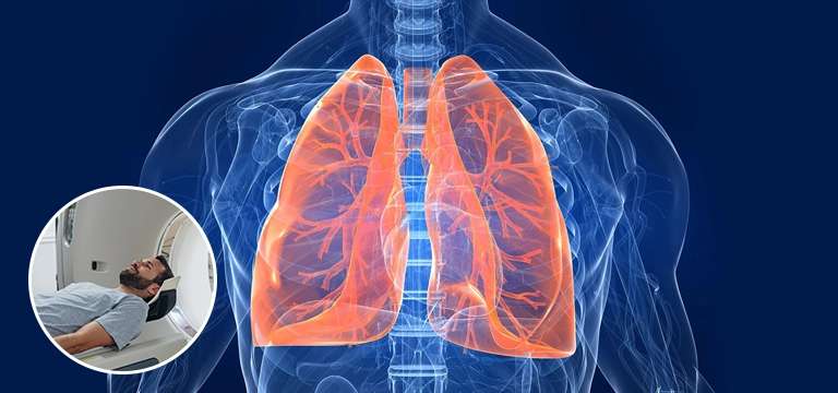
A lung ventilation scan, also known as a lung ventilation/perfusion (V/Q) scan, is a medical imaging test used to assess the airflow in the lungs. It provides information about how well air reaches different areas of the lungs.
What is Lung Ventilation Scan?
A lung ventilation scan, also known as a lung ventilation/perfusion (V/Q) scan, is a medical imaging test used to assess the airflow in the lungs. It provides information about how well air reaches different areas of the lungs. During a ventilation scan, the patient inhales a radioactive gas or aerosol through a mask or mouthpiece. The radioactive material can be inhaled directly or through a breathing apparatus. As the patient breathes, a special camera called a gamma camera is used to take images of the lungs.
The gamma camera detects the radiation emitted by the inhaled material and creates images that show the distribution of air within the lungs. The ventilation scan helps identify areas of the lungs where there may be decreased airflow, blockages, or abnormalities. It can be particularly useful in diagnosing conditions such as chronic obstructive pulmonary disease (COPD), asthma, or assessing lung function prior to surgery.
Why do I need a Lung Ventilation Scan Test?
A lung ventilation scan may be recommended by your healthcare provider for several reasons. Here are some common situations where a lung ventilation scan may be needed:
- Evaluation of Lung Function: If you are experiencing symptoms such as shortness of breath, wheezing, or difficulty breathing, a lung ventilation scan can help assess your lung function. It can provide information about the distribution of air within your lungs and identify any areas of decreased airflow or ventilation abnormalities.
- Respiratory Conditions: If you have a known respiratory condition such as asthma or chronic obstructive pulmonary disease (COPD), a lung ventilation scan can help evaluate the extent and severity of the condition. It can help determine the effectiveness of your current treatment plan or guide adjustments to your medications.
- Preoperative Assessment: Prior to certain surgeries, especially those involving the lungs or thoracic region, a lung ventilation scan may be performed to assess lung function and determine the risk of complications during the procedure. This can help the surgical team make informed decisions about the surgical approach and postoperative care.
- Pulmonary Embolism: A lung ventilation scan, in combination with a perfusion scan, is commonly used to diagnose pulmonary embolism, which is a blood clot in the lung's blood vessels. The scan can help identify areas of the lung with reduced or no blood flow due to blockages, assisting in the diagnosis and management of this potentially life-threatening condition.
- Occupational and Environmental Exposure: If you have been exposed to substances or environments that could potentially affect your lung function, such as industrial pollutants or hazardous chemicals, a lung ventilation scan may be recommended to evaluate the impact on your respiratory system.
- Follow-up After Treatment: If you have received treatment for a lung condition or undergone interventions such as bronchial thermoplasty or lung volume reduction surgery, a lung ventilation scan can be performed to assess the response to treatment and monitor the progress of recovery.
- Lung Disease Progression: For individuals with chronic lung diseases such as interstitial lung disease or pulmonary fibrosis, a lung ventilation scan can help track disease progression and guide treatment decisions.
- Assessment of Lung Transplant Candidates: Before a lung transplant, a comprehensive evaluation of lung function is necessary. A lung ventilation scan can provide valuable information about lung health and help determine if a transplant is suitable.
What happens during a Lung Ventilation test?
During a lung ventilation test, also known as a lung ventilation/perfusion (V/Q) scan, the following steps are typically involved:
- Preparation: Before the test, you will be given specific instructions by the healthcare staff. These instructions may include avoiding certain medications, such as bronchodilators, for a period of time before the test. You may also be asked to refrain from smoking and to wear loose, comfortable clothing.
- Radiopharmaceutical Administration: The test involves the use of a small amount of a radioactive substance that can be inhaled. This substance may be a gas, such as xenon-133, or an aerosol, such as technetium-99m. The radioactive substance is considered safe and will not cause any harm or discomfort.
- Inhalation of the Radiopharmaceutical: You will be positioned in front of a machine that delivers the radioactive substance and may be asked to change positions or lie in different positions to capture images from various angles. This allows for a more comprehensive assessment of lung ventilation. You may wear a mask or mouthpiece connected to the delivery system. As you inhale, the machine will release the radioactive material, and you will be asked to hold your breath for a short time to allow for image acquisition.
- Imaging: After inhaling the radioactive substance, you will be positioned in front of a gamma camera, which is a special imaging device. The camera will take images of your lungs as you breathe in and out. The gamma camera detects the radiation emitted by the radioactive substance and creates images that show the distribution of air within your lungs.
- Image Interpretation: The obtained images will be reviewed and interpreted by a radiologist or a nuclear medicine specialist. They will analyze the images to assess the ventilation patterns in your lungs and identify any abnormalities or areas of decreased airflow.
- Perfusion Scan (optional): In some cases, a perfusion scan may also be performed in conjunction with the ventilation scan. This involves the injection of a radioactive tracer into a vein, and images are taken to evaluate the blood flow to the lungs. The combination of the ventilation and perfusion scans can provide comprehensive information about both airflow and blood flow in the lungs.
- Repeat Imaging: In some cases, the imaging process may be repeated after a certain period of time or after the administration of certain medications. This can help evaluate changes in lung function over time or assess the response to specific interventions.
- Additional Tests: Depending on the specific clinical situation, additional tests or imaging procedures may be performed along with the lung ventilation scan. For example, a chest X-ray or a CT scan may be recommended to provide further information about the structure and condition of the lungs.
What is the price of a Lung Ventilation Scan Test?
A rough estimated price for a lung ventilation scan in India can range from Rs. 5,000 to Rs. 15,000 or more. The cost of a lung ventilation scan can vary depending on several factors, including the healthcare facility, geographical location, additional tests or services included, and individual insurance coverage. It's important to note that these are approximate figures, and the actual cost may differ It is recommended to contact the specific healthcare facility where you plan to have the test performed. They can provide you with the most up-to-date and relevant pricing details based on their services and location.
Best Diagnostic Centre for Lung Ventilation Scan in Delhi?
One of the best Diagnostic Centre for Lung Ventilation Scan in Delhi is Ganesh Diagnostic and Imaging Centre. At Ganesh Diagnostic and Imaging Centre, we are dedicated to providing the highest quality lung ventilation scans in Delhi. Our state-of-the-art facility is equipped with advanced imaging technology and staffed by a team of skilled radiologists and technicians. We prioritize accuracy, efficiency, and patient comfort throughout the scanning process. Whether you require a lung ventilation scan for diagnostic or monitoring purposes, our experienced team ensures precise and timely results. Trust Ganesh Diagnostic and Imaging Centre as the best choice for your lung ventilation scan needs in Delhi, where exceptional care and reliable diagnostics are our top priorities.


.webp)


