MRI (Magnetic Resonance Imaging) of the knee joint is a very useful diagnostic tool that assists in getting detailed visualization and evaluation of the intricate structures of the knee. This is a non-invasive imaging...
Introduction
MRI (Magnetic Resonance Imaging) of the knee joint is a very useful diagnostic tool that assists in getting detailed visualization and evaluation of the intricate structures of the knee. This is a non-invasive imaging technique, which generates valuable information in the bones, cartilage, ligaments, tendons, and other soft tissues. All these constitute the knee joint.
MRI scans work by making use of magnetic fields and radio waves that help in capturing high-resolution images, thereby assisting healthcare professionals and doctors to diagnose and manage various ailments including knee-related conditions.
In this article, we will explore the intricacies of an MRI knee joint, its procedure, common findings, benefits, shortfalls, and frequently asked questions.
MRI Knee Joint: An overview
Before diving into the MRI knee joint, let's first study the complex structures within the knee, which is incidentally the largest joint in the body. It connects the thighbone (femur) to the shinbone (tibia) and includes the kneecap (patella) and various ligaments, tendons, muscles, and cartilage.
The knee joint is responsible for a lot of essential movements like flexion, extension, and rotation, making it prone to injuries and other conditions such as ligament tears, meniscus tears, and arthritis among others. And an MRI plays a pivotal role in assessing and diagnosing these issues related to the knees. MRI of the knee joint provides a detailed visualization aiding in treatment decisions.
MRI Knee Joint: Its Procedure and Preparation
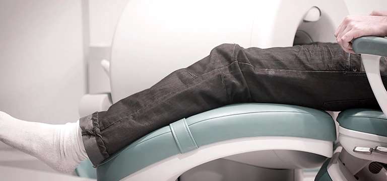
An MRI knee joint procedure usually follows a specific protocol. Before the scan, the patient has to change into a gown the hospital provides and remove any metallic objects in the body, as they can jeopardize the imaging process. The patient then lies flat on a movable table, which slides into the MRI machine. One has to remain still during the MRI scan procedure so that clear and accurate images can be captured.
During the scan, the machine generates strong magnetic fields and radio waves, which cause the body's atoms to emit signals. These signals are captured by the MRI machine and converted into detailed images of the knee joint. Be assured that this process is painless, but the machine may make loud noises. To ensure a patient’s comfort, earplugs or headphones are made available by the staff administering the scan.
In some cases, the use of a contrast agent may become necessary which will be injected intravenously to enhance the visibility of certain structures within the knee joint. This contrast material can provide additional information to assist in the diagnosis.
Common Findings & Interpretation of MRI knee joint
MRI knee joint can reveal a plethora of findings that aid in the diagnosis and evaluation of knee-related conditions. Some of the common findings include ligament tears (such as anterior cruciate ligament or ACL tears), meniscal tears, cartilage damage, tendon injuries (such as patellar tendon or quadriceps tendon tears), joint effusion (excess fluid within the joint), and signs of arthritis or inflammation. You can book arthritis test here.
After that, the images obtained from MRI knee joint scans are then carefully analyzed by radiologists who specialize in musculoskeletal imaging. These experts are trained to interpret the images and provide detailed reports to the referring healthcare provider.
The radiologist thereafter evaluates the condition of the bones, cartilage, ligaments, tendons, and other structures within the knee joint, noting any abnormalities or signs of injury or disease. Their interpretation gives the healthcare provider a path to making an accurate diagnosis and developing an appropriate treatment plan.
Benefits & Limitations of MRI Knee Joint
MRI knee joint offers numerous benefits in the assessment and management of knee-related problems. At the outset, it provides a non-invasive means of obtaining detailed images of the knee joint, eliminating the need for exploratory surgeries. Since it is a non-invasiveness process, it reduces a patient’s discomfort and enables faster recovery times.
Secondly, MRI offers exceptional soft tissue contrast, paving the way for precise evaluation of structures like ligaments, tendons, and cartilage. This detailed visualization aids in accurate diagnosis and helps healthcare professionals to determine the severity and extent of injuries or conditions.
Additionally, an MRI knee joint is highly impactful in diagnosing subtle abnormalities and early signs of degenerative ailments in an individual. It can identify small tears or defects in ligaments, menisci, or cartilage that may not be visible through other imaging modalities.
However, it's worthwhile to mention that an MRI knee joint also has certain limitations. The procedure can be time-consuming, typically taking 30 to 60 minutes to finish. Patients need to remain still during the scan, which can be challenging for those who experience discomfort or have difficulties with immobility.
Furthermore, some patients may experience claustrophobia inside the MRI machine, but healthcare providers can come up with techniques or medications to assuage anxiety in them.
Another challenge to consider here is the cost of an MRI knee joint, which can vary depending on factors such as geographical location, healthcare facility, insurance coverage, and any additional services required. You should consult with your healthcare provider or imaging facility to understand the potential costs associated with the procedure.
MRI knee joint price
Ascertaining the precise rate of an MRI knee joint price can be complex as mentioned above. It is governed by various factors, making it challenging to provide a precise figure.
The cost of an MRI knee joint is finalized based on factors like geographical location, the imaging facility or hospital, the type of insurance coverage, and any additional services or procedures required.
The complexity of the scan and the use of contrast agents, if necessary, may also change the final cost. On top of it, negotiated rates between healthcare providers and insurance companies, as well as deductibles, co-payments, or co-insurance amounts, can further affect the price.
So, you must always contact the specific imaging facility or consult with your insurance provider to get an accurate estimate of the price for an MRI knee joint in your neighborhood.
They can give you detailed information about the associated costs and potential financial assistance programs or payment plans that may be available to help manage the expenses.
It's important to remember that while cost is a consideration, the value and benefits of an MRI knee joint in accurately diagnosing and managing knee-related conditions should also be taken into account.
Conclusion
MRI knee joint is a valuable imaging technique that facilitates detailed examination and evaluation of the complex structures within the knee. It plays a pivotal role in diagnosing and managing various knee-related conditions, helping doctors make informed decisions about treatment plans.
With its non-invasive nature and exceptional soft tissue contrast, the MRI knee joint provides detailed visualization and aids in detecting injuries, tears, and degenerative changes. While it may have certain limitations, such as the need for patient cooperation and potential claustrophobia, the benefits of MRI knee joint scan far outweigh these shortcomings, making it a veritable tool in modern healthcare.
FAQs
Q1. Is an MRI knee joint scan painful?
No, an MRI knee joint scan is painless. But, a few patients may experience mild discomfort from lying still during the scan.
Q2. How long does an MRI knee joint scan take?
The duration of an MRI knee joint scan is typically between 30 and 60 minutes, depending on factors such as the imaging protocol and patient cooperation.
Q3. Can I eat or drink before an MRI knee joint scan?
In most cases you can eat and drink normally before an MRI knee joint scan. However, make sure to follow any specific instructions handed down by your healthcare provider or the imaging facility.
Q4. Will I need an injection during the MRI knee joint scan?
In some cases, a contrast agent may be administered intravenously to enhance the visibility of certain structures within the knee joint. It is not always the case and all MRI knee joint scans may not require contrast material.
Q5. Can I undergo an MRI knee joint scan if I have metal implants?
Generally speaking, individuals with metal implants, such as pacemakers or metal joint replacements, may not be eligible for undergoing MRI scans. However, technological advancements have made it possible for some patients with specific types of implants to undergo MRI scans without any risks. It is important to divulge every detail to your healthcare provider of any implants or metallic objects in your body before scheduling the MRI knee joint.


