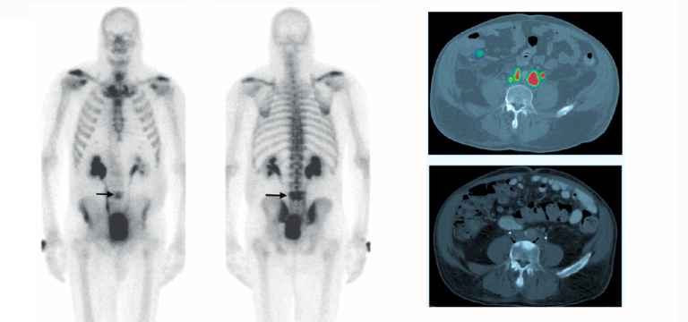
Positron emission tomography (PET) scan refers to the diagnostic imaging modality that detects diseases in a body with the help of a radioactive substance called tracer. The tracer used in the PET scan for bone is F18 fluoride.
Positron emission tomography (PET) scan refers to the diagnostic imaging modality that detects diseases in a body with the help of a radioactive substance called tracer. The tracer used in the PET scan for bone is F18 fluoride. A doctor orders a F18 fluoride PET scan to diagnose metastatic bone disease. It means when the cancer spreads from the organ to the bone.
There are two types of pictures a PET/CT camera can take. The PET scan can show the area where the tracer has collected in the body. The CT (computed tomography) scan offers accurate and detailed pictures of the tissues and structures of the body.
Is a PET scan better than a bone scan?
Each of the scans has its strengths. For instance, bone scans help in diagnosing bony regions in the growth or process. It can indicate a metastatic disease. On the other hand, PET scans can detect irregularities in the body’s biochemical activity, including cells involved in metabolizing glucose extremely fast.
How does a PET/CT scan help in bone scans?
18F-Sodium Fluoride (NaF) bone PET/CT scans is extremely helpful in assisting doctors to get a whole-body view of the skeleton. These images provide great help for the detection and examination of metastatic bone cancer. Some of the most common cancers are mainly associated with metastatic bone disease, prostate, breast, and lung cancer, so, assessing bone metastases is important.
18F-Sodium Fluoride (NaF) PET/CT studies are valuable in:
- Evaluating metastatic bone disease
- Initial staging for patients who are prone to bone metastases
- Situations in which it is important to rule out bone disease to start potentially curative therapy
- Monitoring patients with bone-dominant metastases to check the effectiveness of systemic therapy and exclusion of new metastases at crucial anatomic sites.
For patients 18F-Sodium Fluoride (NaF) PET/CT scans are easy and they offer great comfort and convenience. The duration of time required for scanning is very short that other traditional techniques. Even the waiting period between the scan and the radiopharmaceutical injection is just 45 minutes to an hour for PET/CT scan while it is three hours for conventional studies.
An 18F-Sodium Fluoride (NaF) PET/CT scan is done in 15 to 20 minutes which is a great respite for patients. The other bone scanning techniques need 45 to 60 minutes to complete, which can be a hassle for the patients. Moreover, the complete 18F-Sodium Fluoride (NaF) PET/CT study needs just about an hour, the other studies will require some four hours.
There are many advantages of PET/CT bone scans have over traditional studies, which have been extensively researched in recent times.
Other advantages include of PET/CT scans include:
- A higher accuracy in the detection of both osteolytic and osteoblastic metastases
- Greater level of differentiation between benign versus malignant lesions
- Higher sensitivity compared to 99mTc scans
- Enhanced specificity over other scans
- Increased spatial and contrast resolution
- 18F Sodium Fluoride (NaF) radiopharmaceutical is readily available
- Integration of CT scans provides anatomical data for fusion purposes.
- 3-axis and whole-body viewing akin to FDG PET studies
The procedure
To conduct the PET scan, the technician injects a form of radioactive tracer into the patient.
For a PET scan, the technician injects a form of radioactive sugar, which is also known as FDG into the patient’s blood. Due to the rapid growth of cancer cells within the body, they have a high rate of absorbing sugar. After that, a highly efficient camera captures a picture of areas of radioactivity in the patient’s body. Although the picture is not as precise and detailed as a CT or MRI scan, still it gives useful information about the whole body.
The preferred modality in bone disorders
A total-body PET/CT study, which scans the whole body, can be used to measure all possible osteoporotic bones in a patient.
It is important to note that osteoporosis can impact even the foot and ankle of a patient. So, an entire skeletal scan can offer information on bio-distribution and a better and clear understanding of the osteoporosis process.
In the past few years, several studies have been successfully conducted that quantified osteoporosis in the lumbar spine, hip, tibia, femur, etc. in both humans and animals using NaF PET/CT. Moreover, the most prevalent method of PET/CT imaging is whole-body scanning.
PET scans are also known to properly show the spreading of bone cancer to the lungs, other bones, or for that matter any other parts of the body. PET scans are also invaluable in seeing the efficacy of the treatment in patients.
It is worth mentioning that PET scans can help in detecting cancer earlier than other imaging tests. But certain types of cancer cannot be easily detected on a PET scan. In short, PET scans at times may miss cancers that don’t use a lot of glucose.
Considering a few potential side effects
It is a relief that a PET scan does not entail any pain for the patient, except for the insertion of the IV. The patient might feel a slight cold sensation in the arm when the radiotracer is injected. It is also possible that some people might feel temporary discomfort, redness, or swelling at the injection site.
The patients may feel nervous or anxious if they have:
- Find it difficult to stay still for a long time
- Are afraid of needles
- A fear of enclosed spaces, known as claustrophobia
Conclusion
One of the benefits of the use of PET scan in bone disorders is that it has very low radiation exposure because the radiation usually passes out of the patient’s body in a matter of few hours. One should talk to their consulting doctor if there are any concerns related to radiation exposure during a PET scan.
It is also likely that many doctors will request PET scans in combination with CT scans, MRIs, and other diagnostic modalities because it helps radiologists or specialists in nuclear medicine to evaluate the findings more accurately.
Frequently Asked Questions (FAQs)
Is a PET scan the same as a bone scan?
There are certain differences between bone scans and PET/CT in the detection of bone metastases, which can be attributed to the different mechanisms. A bone scan depends on the osteoblastic response to tumor-induced bone destruction, whereas FDG-PET/CT identifies the metabolic activity of the tumor cells.
What is a bone PET scan?
A PET bone scan is an imaging modality that studies diseases in the body using a radioactive substance called a tracer. The tracer called F18 fluoride is used for a patient’s bone scan. The most common use of PET F18 fluoride bone scans is to detect metastatic bone disease (cancer spreading from an organ to the bone).
Will a PET scan show bone cancer?
PET scans are useful in studying if bone cancer has spread to the lungs, other bones, or other parts of the patient’s body. PET scans are also useful in seeing the response to the treatment in the patient. PET and CT scans can be done by several machines at the same time (PET/CT scan).
Who needs a bone scan?
A regulatory bone scan is very necessary for women aged 65 and older. It is also important for women who have attained the age of 64 or younger but have gone through menopause as they are at a higher risk for osteoporosis.
How long does a PET scan take?
A PET scan usually takes about 15 to 20 minutes. However, the patient has to be in the PET imaging department for about two to three hours. One should ask about any food and drink restrictions the doctor or the technicians present. One should also bring any previous radiology or x-ray images along.
How is a PET scan different from a CT and bone scan?
There are some differences between a PET and a CT scan. One of the main differences between a CT scan and a PET scan is related to their focus. A CT scan is good for creating a detailed non-moving image of bones, organs, and tissues. On the other hand, a PET scan helps doctors to see how the tissues in the body work on a cellular level.
Is there radiation in the bone scan?
A bone scan is done by injecting a very small amount of radioactive material (radiotracer) into a vein, which then traverses through the blood to the bones and organs. It does give off a very little bit of radiation while wearing off. This radiation can be detected by a camera that slowly scans the patient’s body.
Does a PET scan show bone metastases?
Positron emission tomography/computed tomography (PET/CT) has an ability that is inferior to bone scintigraphy (BS) in its accuracy to detect bone metastases despite being a tool for breast cancer (BC) staging.


