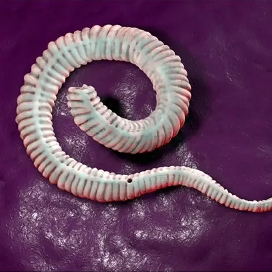
A parasitic nematode that infects people is called the Guinea worm, Dracunculus medinensis. Copepods, which are small crustaceans that carry the worm's contagious larvae, pollute drinking water, which is how it is spread.
A parasitic nematode that infects people is called the Guinea worm, Dracunculus medinensis. Copepods, which are small crustaceans that carry the worm's contagious larvae, pollute drinking water, which is how it is spread.
A painful blister is produced when the female worm, which may reach up to 1 meter in length, migrates through the subcutaneous tissue once the larvae have matured and mated within the body.
The cycle is restarted when the female worm, which normally appears on the lower limbs after roughly a year of infection, emerges from the skin and releases millions of larvae into the water. It can take many weeks for the worm to emerge through the skin, which is a very painful procedure.
Since the 1980s, the World Health Organisation (WHO) has worked to eradicate dracunculiasis, and from an estimated 3.5 million cases in 1986 to just 27 cases in 2020, the disease has been successfully eradicated.
Guinea worm disease: A disease of the past, but with a rich history
Greek and Roman publications, as well as ancient Egyptian medical books from the 16th century BCE, all make mention of the illness.
The Guinea worm has historically been a significant source of illness in many regions of Africa and Asia, and it was especially prevalent in rural communities with subpar sanitation and hygiene. The sickness was frequently tied to droughts, when people were forced to rely on polluted water supplies, and the worm was frequently connected to stagnant water sources including ponds, lakes, and shallow wells.
Early in the 20th century, control techniques including water treatment, health education, and the provision of safe water sources were used to manage and eradicate dracunculiasis.
The World Health Organisation (WHO) established the Global Dracunculiasis Eradication Programme (GDEP) in the 1980s, which marked the beginning of a systematic attempt to eradicate the illness.
Since then, the GDEP has collaborated with local communities, national governments, and NGOs to put into action a variety of interventions, such as water filtering, health education, and active case identification and treatment.
The number of cases has drastically decreased as a result of these efforts, falling from an estimated 3.5 million cases in 1986 to just 27 cases in 2020. Shortly, efforts are being made to guarantee that dracunculiasis is eradicated. The illness is currently seen to be within reach of elimination.
The devil is in the details: A closer look at Dracunculus medinensis
The Morphology of Dracunculus medinensis can be summarized as :
Size
Adult male Guinea worms normally only measure a few centimeters in length, but females can reach lengths of up to one meter.
Body type
The Guinea worm has a long, thin, cylindrical body type. While the back end is tapered, the front end is pointed.
Cuticle
The Guinea worm's exterior surface is coated by a hard, impenetrable cuticle that shields it from the immune system of the host and other external dangers.
Musculature
The Guinea worm has a straightforward muscular structure with muscles that run the length of the body.
Digestive system
The Guinea worm's lack of a functional digestive system allows it to absorb nutrients via its body wall.
Reproductive system
The female Guinea worm is bigger than the male and possesses a long, coiled uterus that may hold up to three million embryos in its reproductive system. For mating, the male possesses a spicule.
Larvae
The Guinea worm's elongated larvae distinctively resemble coils. Through a painful blister on the skin, the mother worm releases them into the sea.
With the help of these morphological characteristics, the Guinea worm can develop, breed, and successfully pass its larvae to copepods in the water, completing its life cycle.
The life cycle of Dracunculus medinensis: A delicate balance between parasite and host
This parasite has an intricate life cycle that includes both people and aquatic creatures. Here are the major elements of its life cycle:
Copepod ingestion
When a human consumes water contaminated with copepods carrying Guinea worm larvae, the Guinea worm life cycle begins.
Larvae release
The larvae break free from the copepods and enter the abdominal cavity through the intestinal wall.
Maturity
The larvae develop into adult male and female Guinea worms over many months, at which point they begin to mate. After mating, the male Guinea worm perishes, but the female Guinea worm keeps growing and moving through the body.
Emergence and release of larvae
The female Guinea worm emerges from the skin, usually on the legs or feet, resulting in a painful blister, after roughly a year of infection. Thousands of larvae are released into the water supply as the blister explodes.
Copepod ingestion
The larvae are consumed by copepods in the water, where they grow to become contagious larvae.
Human infection
The cycle restarts if a human drinks water that contains infected copepods.
Because copepods serve as an intermediary host throughout the larvae's development, the Guinea worm life cycle is unique in that the larvae cannot be passed straight from one person to another. This intricacy makes it difficult to regulate the illness since, to interrupt the cycle, therapies must target both human and animal hosts.
A crippling disease:Dracunculiasis
The Guinea worm, Dracunculus medinensis, is a parasite that causes dracunculiasis, often known as Guinea worm sickness. A painful skin blister, usually on the legs or feet, that develops as a result of this infection can cause impairment and subsequent infections. Here are a few of the infection's salient characteristics:
Incubation period
Approximately one year passes during the infection's incubation phase, during which the human body's Guinea worm larvae mature into adult worms.
Symptoms
A painful skin blister that appears on the skin over several days is the first indication of an infection. Frequent symptoms of the blister include fever, nausea, vomiting, and joint discomfort.
Incapacity
The Guinea worm can result in excruciating pain, incapacity, and loss of movement as it develops and spreads throughout the body. People may find it challenging to work, take care of themselves, or go about their everyday lives as a result.
Secondary infections
When a blister bursts, an open lesion is left behind that might harbor bacteria and develop into cellulitis, abscesses, or sepsis, among other consequences.
Dracunculiasis occurrences have considerably decreased in recent years, but the illness still poses a serious immunoglobulin profile threat to public health in several regions of Africa and Asia. To eradicate Guinea worm disease soon, efforts to manage and eradicate the illness are now undertaken.
A worm in the skin: Clinical diagnosis of Dracunculus medinensis infection-
Here are a few typical diagnostic techniques:
Observation of worm emergence
In regions where dracunculiasis is prevalent, medical professionals may identify the illness by monitoring the appearance of a protruding, painful skin blister that contains a long, slender worm.
History
Medical professionals will often take a thorough medical history and do a physical examination to check for symptoms of the illness, such as blistering, inflammation, and impairment.
Blood testing
Blood tests can be done to check for Guinea worm antibodies, which may signify a current or previous infection.
Imaging tests
Imaging tests including X-rays, ultrasounds, and magnetic resonance imaging (MRI) may be performed to check for signs of the worm in the body or to determine the amount of their presence.
Serological testing
Specific antibodies against the Guinea worm can be found in blood or other body fluids using serological assays, such as the enzyme-linked immunosorbent assay (ELISA).
Polymerase chain reaction (PCR)
PCR is a very sensitive and precise diagnostic method that may be used to amplify and find Guinea worm DNA in blood or tissue samples.
It's crucial to remember that diagnosing dracunculiasis can be difficult, especially in regions where the condition is uncommon or where there are few healthcare services.
Painful but treatable: Treatment of Dracunculiasis
Dracunculus medinensis infections, often known as Guinea worm disease or dracunculiasis, have no recognized treatment or vaccination as of yet. The main treatment includes manually removing the worm from the body, which is a laborious and unpleasant operation. The following are a few typical treatment options:
Water immersion
To relieve the discomfort and help the worm emerge, the injured body area is submerged in cool water.
Extraction of the worm
After it starts to show, medical professionals progressively wind the worm out of the body over a few days or weeks using a stick or another tool. This procedure can be excruciatingly unpleasant and take weeks or months to finish.
Discomfort relief
To assist control the discomfort brought on by the infection, non-steroidal anti-inflammatory medicines (NSAIDs) or opioids may be administered.
Antibiotics
Due to the open wound that the worm's emergence might cause, antibiotics may be administered to prevent or cure subsequent infections.
Wound care
Proper wound care can help avoid secondary infections and speed up recovery. This includes routine cleaning and dressing changes.
Education and preventive programs that promote the use of clean drinking water, proper hygiene and sanitation habits, and the management of the intermediate host (copepods) can aid in the prevention of new infections.
No More Worms : Let's Fight Dracunculiasis and Win.


