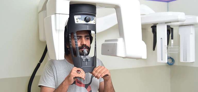
An OPG (Orthopantomogram) X-ray is a specialized dental imaging technique that provides a panoramic or wide-angle view of the entire upper and lower jaws, including the teeth, jawbones, and surrounding structures.
What is OPG X-Ray?
An OPG (Orthopantomogram) X-ray is a specialized dental imaging technique that provides a panoramic or wide-angle view of the entire upper and lower jaws, including the teeth, jawbones, and surrounding structures. It is also known as a panoramic dental X-ray. During an OPG X-ray, the patient's head is positioned in a machine that rotates around the head, capturing a comprehensive image of the oral region. The machine emits X-rays, which pass through the patient's oral structures and are detected by a film or digital sensor.
The resulting image shows a detailed view of the teeth, their positioning, and the overall condition of the dental structures. It allows dentists to assess dental health, detect abnormalities or dental pathologies, evaluate the position of teeth, and plan various dental treatments, such as orthodontics, implants, extractions, or oral surgeries. OPG X-rays are valuable diagnostic tools, providing a broader perspective compared to traditional intraoral X-rays. They aid in diagnosing dental issues that may not be visible through a regular dental examination alone. OPG X-rays are safe, relatively quick, and offer numerous benefits in dental assessment, treatment planning, patient education, and the early detection of dental and jawbone abnormalities.
OPG Preparation & Procedure
Preparing for an OPG (Orthopantomogram) X-ray is relatively straightforward. Here's an overview of the preparation and procedure:
- Preparation:
- Clothing: The patient can wear his/her regular clothing for an OPG X-ray. However, it is essential to remove any metallic objects, such as jewellery, eyeglasses, or hair accessories, as they can interfere with the imaging process.
- Informing the technologist: Before the procedure, inform the technologist if the patient is pregnant or if the patient has any dental appliances, such as braces or dentures. These details can help ensure the proper adjustments are made during the imaging process.
- The OPG X-ray Procedure:
- Positioning: The patient will be asked to stand or sit upright next to the OPG machine. The technologist will position the patient head in the machine's headrest and adjust it for optimal alignment.
- Bite position: The patient will be instructed to bite gently on a plastic bite stick or a bite guide. This helps to ensure that the patient’s teeth are in the correct position for the panoramic image.
- Staying still: During the X-ray, it is essential to remain still and follow the technologist's instructions. Movement can result in blurry images, and the procedure may need to be repeated.
- Machine rotation: The machine will rotate around the head of the patient, capturing a panoramic image of the entire upper and lower jaws. It typically takes a few seconds for the machine to complete its rotation.
- Completion: Once the imaging is complete, the technologist will review the images for clarity and make sure all necessary views are captured. The patient will be informed that the procedure is finished, and the patient can resume his/her normal activities.
The images obtained from the OPG X-ray will be analysed by a dentist or dental specialist. They will assess the condition of the teeth, jawbones, and surrounding structures, and use the information to evaluate the patient’s dental health, plan treatments, and address any concerns or issues.
Benefits of an OPG X-Ray
OPG (Orthopantomogram) X-rays offer several benefits in dental imaging. Here are some of the advantages:
- Non-invasive procedure: OPG X-rays are non-invasive and do not require any injections or incisions. They are performed externally, without any discomfort or pain to the patient.
- Comprehensive view: OPG X-rays provide a panoramic or wide-angle view of the entire upper and lower jaws in a single image. This allows dentists to assess the overall dental and skeletal structures, including teeth, jawbones, sinuses, and temporomandibular joints (TMJ).
- Dental assessment: OPG X-rays help dentists evaluate the condition and position of teeth, identify dental abnormalities (such as impacted or supernumerary teeth), and assess the presence of dental caries (cavities) or periodontal disease. It aids in diagnosing dental issues that may not be visible through a clinical examination alone.
- Treatment planning: OPG X-rays assist in planning various dental treatments, including orthodontics, dental implants, extractions, and oral surgeries. The comprehensive image allows dentists to assess the feasibility, positioning, and alignment of dental procedures accurately.
- Cost-effective for dental professionals: OPG X-rays can be cost-effective for dental professionals, as they provide a comprehensive assessment of the oral structures in a single image. This eliminates the need for multiple intraoral X-rays and reduces overall costs associated with imaging.
- Pathology detection: OPG X-rays can detect and evaluate dental and jawbone pathologies, such as cysts, tumours, infections, or fractures. It helps in early identification and appropriate management of these conditions.
- Time-saving and cost-effective: Compared to traditional intraoral X-rays, which capture images of individual teeth, an OPG X-ray captures the entire oral region in one scan. This saves time and reduces the need for multiple X-ray exposures, making it a cost-effective option.
- Patient education: OPG X-rays provide visual aids for patient education. Dentists can show patients their panoramic images, explaining dental conditions, treatment options, and potential areas of concern, promoting better understanding and informed decision-making.
- Temporomandibular joint (TMJ) evaluation: OPG X-rays can provide valuable information about the temporomandibular joints, helping in the assessment of TMJ disorders, jaw alignment, and occlusion (bite).
What happens during an OPG?
During an OPG (Orthopantomogram), the following steps typically occur:
- Positioning: The patient will be asked to stand or sit in front of the OPG machine. The technologist will position the patient’s head against a headrest to ensure stability and proper alignment.
- Bite position: The patient will be provided with a plastic bite stick or a bite guide to bite on gently. This helps align the patient’s teeth and jaws in the correct position for the panoramic image.
- Staying still: It is crucial to remain as still as possible during the OPG scan. The technologist will instruct the patient to keep the head and body still throughout the procedure. Any movement may affect the clarity of the image.
- Machine rotation: The OPG machine will rotate around the head in a semi-circular motion, capturing a panoramic image of the upper and lower jaws. The patient will be asked to hold the patient position and avoid swallowing or talking during the rotation.
- Image capture: As the machine rotates, it emits X-rays to create the image. The X-ray detector or film within the machine captures the X-rays that pass through the oral structures, generating a comprehensive view of the teeth, jawbones, and surrounding structures.
- Completion: Once the rotation is complete, the technologist will review the images obtained to ensure their quality and accuracy. The entire process is relatively quick, typically taking less than a minute.
- After the OPG, the images will be examined by a dentist or dental specialist. They will interpret the images, assess the dental health, identify any abnormalities or concerns, and develop a treatment plan if necessary. They may discuss the findings with the patient, explain any dental issues, and provide recommendations or further instructions based on the results of the OPG.
It's important to note that an OPG is a non-invasive and painless procedure. The level of radiation exposure is low, and the benefits of obtaining a comprehensive view of the patient’s oral structures generally outweigh any potential risks.
Price of OPG Scan
The cost of an OPG (Orthopantomogram) X-ray in India can vary depending on factors such as the city, type of facility, and whether it is done at a government hospital, private hospital, or diagnostic centre. Additionally, prices may differ based on the specific dental clinic or radiology centre the patient visit. On average, the cost of an OPG X-ray in India can range from approximately INR 300 to INR 1,500. However, it's important to note that these figures are approximate and can vary significantly. The actual cost may be higher or lower depending on the factors mentioned above.
Furthermore, it's worth considering that certain government healthcare programs, insurance plans, or dental clinics may offer OPG X-rays at subsidized rates or as part of a package of dental services.


