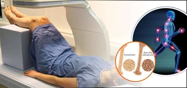
Bone mass density, also known as bone mineral density (BMD), is a measure of the amount of mineral content in bone tissue. It is an important indicator of bone health and strength.
Bone mass density, also known as bone mineral density (BMD), is a measure of the amount of mineral content in bone tissue. It is an important indicator of bone health and strength.
BMD is typically measured using a specialized test called dual-energy X-ray absorptiometry (DXA or DEXA). This test involves passing low-dose X-rays through bones, usually the hip and spine, to determine the amount of mineral present. The results are then compared to reference values to determine the individual's bone health status.
The measurement of BMD provides information about bone strength and the risk of fractures. A higher BMD indicates stronger bones, while a lower BMD suggests reduced bone density and increased vulnerability to fractures. BMD is commonly used to diagnose osteoporosis, a condition characterized by low bone mass and deterioration of bone tissue, which can lead to increased fracture risk.
BMD is influenced by various factors, including genetics, age, sex, hormonal status, lifestyle factors (such as diet and exercise), and certain medical conditions or medications. Regular monitoring of BMD can help in assessing bone health, guiding treatment decisions, and evaluating the effectiveness of interventions aimed at improving bone density and reducing fracture risk.
When Does Bone Mass Density Start Decreasing?
Bone mass density (BMD) typically starts to decrease in both men and women as they age. However, the rate of decline may vary among individuals and can be influenced by several factors.
In women, BMD loss tends to accelerate after menopause. During menopause, there is a significant decrease in estrogen levels, which plays a crucial role in maintaining bone density. This hormonal change leads to an increased rate of bone loss, making women more susceptible to osteoporosis and fractures.
In men, BMD decline occurs more gradually compared to women. Testosterone, the primary male sex hormone, helps maintain bone health in men. As men age, testosterone levels gradually decrease, resulting in a gradual decline in BMD.
While age and hormonal changes are major contributors to BMD decline, other factors can also affect bone health. These include:
- Lifestyle factors: A sedentary lifestyle, lack of weight-bearing exercises, poor nutrition (especially low calcium and vitamin D intake), smoking, excessive alcohol consumption, and inadequate sun exposure can contribute to decreased BMD.
- Medical conditions: Certain medical conditions such as hyperthyroidism, hypogonadism, celiac disease, rheumatoid arthritis, and chronic kidney or liver disease can impact bone health and accelerate BMD loss.
- Medications: Some medications, such as glucocorticoids (steroids), anticonvulsants, certain cancer treatments, and long-term use of proton pump inhibitors, can negatively affect bone density.
It's important to note that while BMD naturally declines with age, the rate and severity of bone loss can be influenced and mitigated by lifestyle choices and appropriate medical interventions. Engaging in regular weight-bearing exercises, consuming a balanced diet rich in calcium and vitamin D, avoiding smoking and excessive alcohol consumption, and discussing bone health with a healthcare professional can help maintain optimal bone density and reduce the risk of fractures.
What is a Dexa Scan?
A BMD DXA scan, also known as a bone mineral density dual-energy X-ray absorptiometry scan, is a specialized imaging test used to measure bone density. It is considered the gold standard for assessing bone health and diagnosing conditions like osteoporosis.
During a DXA scan, the patient lies on a padded table while a machine passes two low-dose X-ray beams through the bones being examined, typically the hip and spine. The machine measures the amount of X-ray energy that passes through the bone, and based on the differences in absorption, it calculates the bone mineral density.
DXA scans provide two important measurements:
- T-Score: This score compares an individual's bone density to that of a healthy young adult of the same sex. It is used to diagnose osteoporosis and determine the risk of fractures. A T-score of -1.0 or above is considered normal, between -1.0 and -2.5 indicates low bone mass (osteopenia), and -2.5 or below indicates osteoporosis.
- Z-Score: This score compares an individual's bone density to that of an average person of the same age, sex, and size. It helps identify factors other than aging that may be contributing to bone loss, such as medical conditions or medications.
The DXA scan is a quick and painless procedure, typically taking around 10 to 30 minutes to complete. The radiation exposure is very low, equivalent to or even less than a standard chest X-ray.
The results of a DXA scan provide valuable information about bone health and fracture risk. They can help guide treatment decisions, monitor the effectiveness of interventions, and track changes in bone density over time. It is advisable to discuss the results with a healthcare professional who can interpret them and provide appropriate recommendations based on the individual's specific circumstances.
What Are the indications of DXA bone density test?
A DXA bone density test, also known as a BMD (bone mineral density) test, is typically indicated in the following situations:
- Osteoporosis screening: DXA scans are commonly used to screen for osteoporosis, especially in postmenopausal women and older adults. The test helps identify individuals with low bone density and assess their risk of fractures.
- Fracture risk assessment: DXA scans are used to evaluate the risk of fractures in individuals who have not yet experienced a fracture but may have risk factors such as age, family history, certain medical conditions, or long-term use of medications that can affect bone health.
- Monitoring bone health: Individuals who have been diagnosed with osteoporosis or low bone mass may undergo DXA scans at regular intervals to monitor changes in bone density and assess the effectiveness of treatment interventions.
- Evaluation of treatment effectiveness: DXA scans can help determine whether prescribed treatments for osteoporosis, such as medication, lifestyle modifications, or hormone replacement therapy, are improving bone density and reducing fracture risk.
- Assessment of certain medical conditions or medications: Some medical conditions, such as hyperthyroidism, hypogonadism, rheumatoid arthritis, or chronic kidney disease, can affect bone health. DXA scans may be ordered to assess bone density in individuals with these conditions. Additionally, certain medications, such as long-term glucocorticoid (steroid) use, can contribute to bone loss, and DXA scans can help monitor bone density in individuals taking these medications.
- Preoperative evaluation: DXA scans may be used as part of preoperative evaluations, especially for individuals undergoing spinal surgery or other procedures where bone density may impact surgical planning or risk assessment.
What are The Contraindications of Dexa test?
Dual-energy X-ray absorptiometry (DXA) scans, also known as DEXA scans, are generally safe and well-tolerated. However, there are a few contraindications and considerations to be aware of:
- Pregnancy: DXA scans involve a small amount of radiation exposure. While the radiation dose is generally considered safe for adults, it is recommended to avoid DXA scans during pregnancy unless the potential benefits outweigh the risks. If a DXA scan is deemed necessary during pregnancy, appropriate radiation safety measures should be taken.
- Metal objects: Metal objects, such as jewelry, belts, zippers, or clothing with metal buttons, may interfere with the DXA scan and affect the accuracy of the results. It is important to remove or avoid wearing any metal objects in the area being scanned.
- Recent contrast agent injection or nuclear medicine scans: Certain contrast agents used for diagnostic imaging, such as those containing barium or iodine, can interfere with DXA scan results. It is generally recommended to wait for a specific period, usually 7-14 days, after receiving a contrast agent or undergoing a nuclear medicine scan before performing a DXA scan.
- Severe obesity: DXA scans may have limitations in individuals with severe obesity. The size and weight of the individual may exceed the capacity of the DXA scan table or limit the accuracy of the measurements. In such cases, alternative methods or specialized equipment may be considered.
It is important to inform the healthcare provider performing the DXA scan about any existing medical conditions, previous contrast agent injections, pregnancy, or the presence of metal objects to ensure appropriate precautions are taken or alternative imaging options are considered if necessary.
Overall, DXA scans are considered safe and the benefits of obtaining valuable information about bone health and fracture risk generally outweigh the risks associated with the minimal radiation exposure involved.
Conclusion
In conclusion, bone mass density (BMD) is a crucial measure of bone health and strength. It serves as an indicator of the amount of mineral content present in bone tissue. BMD decreases naturally as individuals age, with women experiencing an accelerated decline after menopause. Various factors, such as genetics, hormonal status, lifestyle choices, and certain medical conditions or medications, can influence BMD. Regular monitoring of BMD through specialized tests like DXA scans helps diagnose osteoporosis, assess fracture risk, evaluate treatment effectiveness, and guide interventions to improve bone density. While DXA scans are generally safe and well-tolerated, precautions should be taken for individuals who are pregnant, have recently received contrast agents, or have severe obesity. Following specific instructions provided by healthcare providers or imaging centers ensures optimal preparation for the scan. By staying proactive about bone health and working closely with healthcare professionals, individuals can take steps to maintain and improve their bone density, thereby reducing the risk of fractures and promoting overall well-being.


