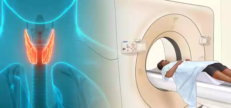
Located in front of the neck, the thyroid gland is a small gland in the human body, which is in the shape of a butterfly. It is one of the most important glands in the body because it secretes those hormones which are very...
Introduction
Located in front of the neck, the thyroid gland is a small gland in the human body, which is in the shape of a butterfly. It is one of the most important glands in the body because it secretes those hormones which are very crucial for regulating metabolic functions. The thyroid has an indirect sway over the functioning of the whole body and development. But, thyroid ailments are also pretty common, especially among women.
There is thyroid cancer afflicting the thyroid gland, although it is rare. But it is also seen as the most common endocrine malignancy. Just like thyroid diseases, even thyroid cancer is most commonly found in females. The respite is that, unlike other cancers, thyroid cancer is known to have the best prognosis and is treatable with successful diagnosis and timely intervention.
Symptoms of Thyroid Cancer
As a disease, thyroid cancer develops very slowly and hence it is generally seen that in the early stages, it does not exhibit any signs and symptoms. As the disease progresses, it may show up the following signs and symptoms in patients:
- A lump in front of the neck or on either side of the throat, which can be felt
- Pain while swallowing
- Difficulty in talking or breathing
- Sudden hoarseness and change in the tone of voice
- Persistent coughing
Risk Factors Linked to Thyroid Cancer
The risk factors attributed to developing Thyroid cancer have always been debatable. The disease is thought to be developing as a consequence of certain cellular changes due to mutation of the genetic material. However, certain factors are widely accepted as its causes are:
- Women are at a greater risk of developing thyroid cancer. It is because of the role of the estrogen hormone present in females, which is suggested by several studies.
- High level of radiation exposure.
- Hereditary genetic syndromes: Certain genetic syndromes are considered very risky for the development of thyroid carcinoma. Also, certain thyroid cancer is known to be in the genes.
- Deficiency in the iodine level can also contribute to the risk of thyroid cancer in people.
Different Types of Thyroid Cancer
To determine the different types of thyroid cancer it is required to conduct a pathological examination of a tissue sample from the thyroid. Further, the different types influence the prognosis and treatment plan.
The different types of thyroid cancer are papillary, follicular, medullary, anaplastic, hurthle cell, and Lymphoma.
Diagnostic Techniques for Thyroid Cancer
Different diagnostic techniques for thyroid cancer are as below:
Physical examination & medical history: It is the initial step to access in detail the medical history and physical evaluation of the lumps including other signs & symptoms.
Blood Tests: Although blood tests are not capable to diagnose thyroid cancer, they are still used to know more about the functions of the thyroid gland. Most commonly assessed in the tests include thyroid-stimulating hormones, thyroglobulin, T3 and T4 hormones, calcitonin, and carcinoembryonic antigen.
A very high level of thyroglobulin is regarded as an indicator after surgery, pointing to the presence of recurring cancer cells or residual cancer. Thyroglobulin is produced by the thyroid tissues, so when the thyroid gland is surgically removed, the thyroglobulin level is expected to be low.
Imaging Tests: Various imaging modalities such as ultrasound, X-ray, magnetic resonance imaging (MRI), computed tomography (CT), and positron emission tomography (PET) are extensively used to diagnose thyroid cancer and also as an assessment of its spread.
Radioiodine Scans: During this test, a small amount of radioactive iodine is administered to patients, and specialized gamma camera images are captured. Thyroid cells have a propensity to get attracted to iodine, facilitating thyroid tissue located anywhere in the body to absorb this iodine. This technique can be used for all types of thyroid cancer except for the medullary type, as they exhibit a significantly low affinity to iodine.
Biopsy: It can confirm the diagnosis and also the type of thyroid cancer.
PET-CT Scan and thyroid cancer diagnosis
PET-CT scanning helps assess the molecular functioning of tissues, using a radioactive substance F-18 fluorodeoxyglucose (FDG), which is similar to glucose molecules. This radioactive material gathers in different tissues throughout the body based on their metabolic activity. Due to their rapid division and exponential growth, tumor cells exhibit higher metabolic activity and as a result, attract glucose at a higher rate. Hence, there is an increased uptake of the radioactive material, leading to a conspicuous bright spot in the PET scan.
While FDG is the commonly used radiotracer in PET scans, novel tracers such as Iodine-124, 18F-DOPA, Iodine-131, or Iodine-123 are being assessed for their potential application in thyroid malignancies.
Iodine-123:
- High image quality
- Costly
- Short half-life
- Restricted availability
Iodine-131:
- Cheaper
- Easily available
- Inferior resolution images
Iodine-124:
- Good half-life
- High sensitivity
- Better-resolution images
- Highly sensitive to detecting recurrence
Several studies have 124I PET-CT to be comparable related to sensitivity and usefulness to FDG PET-CT.
Role of PET-CT in Thyroid Cancer
PET scans have brought about a paradigm shift in the field of cancer diagnostics including Thyroid cancer. The radioactive material used in PET scans is injected into the body of the patient through a vein and thereafter specialized cameras take the images. PET scans are extremely helpful in determining how the organs and tissues are working based on the biochemical and metabolic activities happening in the tissues.
Thus, it is by studying the changes in the tissue metabolic activity doctors can identify the disease earlier, much before any occurrences of structural or anatomic changes, which may be detected only later in an imaging procedure.
When combined, a PET scan and CT imaging can provide better cancer diagnosis and detection of its spread. While the PET scan offers insights into the functional aspects of tissues, the CT scan is useful in providing anatomical and morphological information about any abnormalities. Hence, a combined PET-CT scan delivers dual diagnostic clarity, improving the accuracy and efficacy of the diagnosis process.
Although PET-CT may not e a primary diagnostic tool in thyroid cancer, it plays a great role as a prognostic indicator apart from recurrent thyroid cancer cases.
Conclusion
Thyroid cancer is known to have a good prognosis and is fully curable. Even recurrent thyroid cancer cases are better than other cases when it comes to prognosis. We have seen that despite not being the main diagnostic modality in thyroid cancer, PET-CT has proven to be the best prognostic technique and a crucial follow-up tool in thyroid cancer patients.
FAQs
Do thyroid nodules show on PET scans?
Among common imaging techniques, PET scans can identify thyroid nodules when looking for other cancers. It is concerning for cancer if nodules/masses are PET-positive. During the diagnosis or staging of non-thyroid cancers, approximately 1-2% of PET scans reveal unexpected detection of thyroid lesions.
What cancers can't be detected by PET scan?
A negative PET scan may fail to detect cancerous tumors, such as a type of lung cancer (bronchioalveolar carcinomas), tumors that grow from neuroendocrine cells (carcinoid tumors), low-grade lymphomas, etc.
What is the first test for thyroid cancer?
The first test for thyroid cancer is an ultrasound exam when the doctor notices a lump or swelling in the neck. Thyroid nodules are also often visible on CT scans conducted for another reason. Doctors may also order blood tests to check the patient’s thyroid hormone levels.
Can a PET scan tell if a nodule is cancerous?
When a nodule actively grows or exhibits high metabolic activity, it will be visually highlighted in a PET (positron emission tomography) scan. The intensity of the nodule's brightness on the PET scan directly correlates with its likelihood of being cancerous. Moreover, the PET scan examines the entire body and can determine if the cancer has metastasized or spread to other areas.
Does PET scan show malignancy?
A PET scan is very useful to find a malignant (cancerous) tumor or whether it is benign (not cancerous). It is different from other imaging tests such as CT or MRI that show anatomy. The PET scan deals with physiological changes and cellular activity, so cancer may be detected much earlier.
Why PET scan is not recommended?
It is not recommended for people who are pregnant, breastfeeding, or chest feeding. The radiation in a PET scan can harm the fetus and it can pass to an infant in breast milk. Also, those who have an allergic reaction may get affected by PET scan radioactive tracers or CT scan contrast dyes. However, these allergic reactions are usually mild and extremely rare.
What cancers show up on a PET scan?
PET scans are very helpful in detecting cancer and the extent of its spread. It can also show solid tumors in the brain, thyroid, prostate, lungs, and cervix. PET scans can also evaluate the occurrence of colorectal, melanoma, lymphoma, and pancreatic tumors.
Is a PET scan Painful?
A PET-CT scan is not painful. But for a few patients, some positions might be uneasy or tiring.


