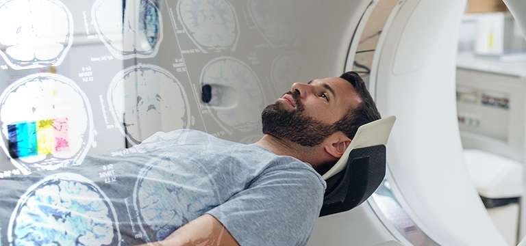
During a nuclear medicine scan, a radiotracer is administered to the patient either by injection, inhalation, or ingestion. The radiotracer emits gamma rays, which are detected by a special camera called a gamma camera or a...
A nuclear medicine scan is a medical imaging technique that uses small amounts of radioactive materials, called radiotracers or radiopharmaceuticals, to diagnose and treat diseases. It provides valuable information about the structure and function of organs and tissues within the body.
During a nuclear medicine scan, a radiotracer is administered to the patient either by injection, inhalation, or ingestion. The radiotracer emits gamma rays, which are detected by a special camera called a gamma camera or a positron emission tomography (PET) scanner. These cameras are able to detect the radiation emitted by the radiotracer and create images that show the distribution of the radiotracer within the body.
The images produced by nuclear medicine scans can help in the diagnosis and evaluation of a variety of conditions. They can show how organs are functioning, such as the heart's blood flow or the brain's metabolic activity. Nuclear medicine scans are commonly used to detect and diagnose conditions such as cancer, heart diseases, thyroid disorders, bone abnormalities, and kidney problems, among others.
In addition to diagnostic purposes, nuclear medicine scans can also be used for therapeutic procedures. For example, radioactive iodine can be used to treat certain types of thyroid cancer, and radiopharmaceuticals can be employed to relieve pain in cancer patients or treat certain forms of hyperthyroidism.
It's important to note that the radiation exposure from nuclear medicine scans is generally considered safe and well-controlled, as the radiotracers used have short half-lives and low levels of radiation. The benefits of the diagnostic or therapeutic information obtained from these scans typically outweigh the risks associated with the radiation exposure. However, as with any medical procedure, the potential risks and benefits should be discussed with your healthcare provider.
Type of Nuclear Medicine Scan
There are various types of nuclear medicine scans, each designed to examine different organs or systems within the body. Here are some common types of nuclear medicine scans:
- Single-Photon Emission Computed Tomography (SPECT): SPECT scans involve the use of a gamma camera that rotates around the patient, capturing multiple images from different angles. It provides three-dimensional images of organs and their function. SPECT scans are commonly used for cardiac imaging, bone scans, and brain imaging.
- Positron Emission Tomography (PET): PET scans use a radiotracer that emits positrons, which are detected by a PET scanner. This scan provides information about metabolic activity in organs and tissues. PET scans are frequently used in oncology to detect and stage cancers, evaluate treatment response, and monitor disease progression. They can also be used to assess brain function and diagnose certain neurological disorders.
- Thyroid Scan: This scan evaluates the structure and function of the thyroid gland. A radiotracer containing radioactive iodine is administered, and the gamma camera captures images of the thyroid gland to assess its size, shape, and function. Thyroid scans are used to diagnose thyroid nodules, hyperthyroidism, and thyroid cancer.
- Bone Scan: This scan is used to evaluate bone conditions and abnormalities, such as bone infections, fractures, bone tumors, and metastatic cancer. A radiotracer is injected into the bloodstream, and the gamma camera captures images of the skeleton, highlighting areas of increased or decreased bone activity.
- Cardiac Stress Test: This scan combines a nuclear medicine study with a stress test to evaluate the blood flow to the heart. A radiotracer is injected during exercise or pharmacological stress, and images are obtained to assess the blood flow to the heart muscle. It helps in diagnosing coronary artery disease and determining the presence of ischemia or areas with reduced blood flow to the heart.
- Renal Scan: Renal scans assess the structure and function of the kidneys. A radiotracer is injected, and images are taken to evaluate kidney blood flow, filtration, and excretion. Renal scans are used to diagnose kidney diseases, assess kidney function, and detect abnormalities like obstructions or renal artery stenosis.
Why do I need a Nuclear Medicine Scan Test?
There are several reasons why your healthcare provider may recommend a nuclear medicine scan. Here are some common situations where a nuclear medicine scan may be necessary:
- Diagnosis: Nuclear medicine scans can provide valuable diagnostic information that may not be obtained through other imaging techniques. They can help identify and characterize diseases or abnormalities in various organs and tissues. For example, they can be used to detect and stage cancers, evaluate cardiac function, diagnose thyroid disorders, assess bone conditions, and identify kidney abnormalities.
- Treatment Planning and Monitoring: Nuclear medicine scans can assist in treatment planning and monitoring the effectiveness of therapies. They can help determine the extent of a disease, evaluate response to treatment, and guide further interventions. For instance, in cancer treatment, nuclear medicine scans can be used to assess tumor response to chemotherapy or radiation therapy.
- Functional Assessment: Nuclear medicine scans provide information about the function and physiology of organs and tissues. They can assess blood flow, metabolic activity, and other functional aspects. These scans are particularly useful in evaluating cardiac function, brain activity, lung ventilation and perfusion, and renal function.
- Disease Progression Monitoring: Nuclear medicine scans can be used to monitor the progression of certain diseases over time. By comparing images taken at different points, healthcare providers can assess the progression or regression of diseases and adjust treatment plans accordingly.
- Preoperative Planning: Nuclear medicine scans can aid in preoperative planning by providing detailed information about the location, extent, and activity of a disease. This helps surgeons in determining the surgical approach, identifying the precise areas to be treated, and minimizing damage to healthy tissues.
- Safety and Efficacy Assessments: Nuclear medicine scans can be utilized to evaluate the safety and efficacy of certain treatments. For example, in nuclear medicine therapy, such as radioactive iodine treatment for thyroid cancer or radioembolization for liver cancer, scans can be performed to confirm the uptake of the radiotracer and assess treatment response.
What happens during a Nuclear Medicine test?
During a nuclear medicine test, here's what typically happens:
- Radiotracer Administration: The process begins with the administration of a radiotracer, which is a small amount of radioactive material. The radiotracer may be injected into a vein, inhaled as a gas, or ingested orally, depending on the specific test being performed and the organ or system being studied.
- Uptake Time: After the radiotracer is administered, there is usually a waiting period called the uptake time. This allows the radiotracer to distribute and accumulate in the target organ or tissue, depending on its specific properties and the metabolic processes being investigated. The waiting time can range from minutes to hours, depending on the test.
- Imaging Procedure: Once the radiotracer has had sufficient time to distribute within the body, the imaging procedure begins. You will be positioned on a scanning table, and a specialized camera, such as a gamma camera or PET scanner, will be used to capture images.
- Gamma Camera: For tests using a gamma camera, the camera will be positioned over or around the area of interest. You may be asked to remain still or change positions during the scan. The camera will detect the gamma rays emitted by the radiotracer, and multiple images will be taken from different angles to create a three-dimensional representation of the organ or tissue being studied.
- PET Scanner: If a PET scan is being performed, you will be positioned on the scanner bed, which will move through a large circular opening in the PET scanner. The PET scanner detects the positrons emitted by the radiotracer, and a computer creates detailed images of the metabolic activity in the area being studied.
- Duration of Imaging: The imaging process typically takes anywhere from a few minutes to an hour, depending on the specific test and the area being studied. You will be asked to remain still during the image acquisition to ensure clear and accurate images.
- Completion of the Test: Once the imaging is complete, you will be able to leave the imaging area unless further imaging or scans are required. The radiotracer administered will naturally lose its radioactivity over time, and it will eventually be eliminated from your body through natural bodily functions, such as urination or bowel movements.
It's important to note that the actual procedure may vary depending on the specific nuclear medicine test being performed and the protocols followed by the healthcare facility.
What is price of the Nuclear Medicine Scan Test?
The cost of a nuclear medicine scan can vary depending on various factors, including the specific type of scan, the healthcare facility, the location, and any additional services or consultations involved.
To obtain accurate and up-to-date pricing information for a nuclear medicine scan in Delhi, you can contact healthcare facilities or hospitals in your area. They will be able to provide you with the most relevant and current pricing details based on your specific requirements and circumstances. You may also consider checking with medical insurance providers to determine if the scan is covered under your insurance plan, as this can affect the overall cost.
Best Diagnostic Centre for Nuclear Medicine Scan in Delhi
Ganesh Diagnostic & Imaging Centre is a highly regarded diagnostic facility in Delhi that offers a wide range of nuclear medicine tests. Renowned for its commitment to delivering comprehensive diagnostic services and exceptional patient care, the centre boasts advanced technology and state-of-the-art facilities to ensure precise and dependable test results. Their team of experienced radiologists and healthcare professionals specialize in nuclear medicine diagnostics and possess extensive expertise in conducting these tests. The centre prioritizes patient comfort, maintaining a welcoming environment, and employs compassionate and knowledgeable staff members who strive to create a positive experience throughout the testing process.


