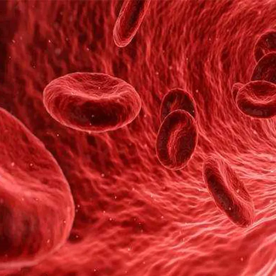
A state of a low extracellular fluid volume known as hypovolemia is typically caused by coupled salt and water loss. To maintain homeostasis, all living things must keep their fluid balances in check. With about 50% to 60% of...
Fluids Matter: Don't Let Hypovolemia Take Control."
Introduction
A state of a low extracellular fluid volume known as hypovolemia is typically caused by coupled salt and water loss. To maintain homeostasis, all living things must keep their fluid balances in check. With about 50% to 60% of the total weight, water is the most prevalent fluid in the body. The extracellular fluid (ECF), which makes up around 25–45% of total body water, and the intracellular fluid (ICF), which makes up 55%–75% of total body water, are further split. The intravascular and extravascular (interstitial) areas make up the remainder of the ECF. ECF is the component that can be measured more easily because arterial blood pressure may be used to estimate it.
When the body loses too much fluid, including water and electrolytes like sodium and potassium, it develops hypovolemia, often referred to as volume depletion. It can occur for a number of reasons, including extreme perspiration, vomiting, diarrhoea, severe burns, or overindulgent urination.
It becomes harder for the heart to pump enough blood to your body when there is significant fluid loss. Organ failure might result from hypovolemic shock when the fluid loss rises. This needs emergency medical care right away.
The Prevalence and Incidence of Hypovolemia: An Epidemiological Perspective
It is challenging to estimate the prevalence of hypovolemia in the general population. One of the most prevalent signs in the acutely unwell patient is hypovolemia. fluid changes, stress, and other etiologies are more frequent in critically ill patients who need intensive care. Overall, there are numerous demographic, environmental, and health-related factors that have a role in the epidemiology of hypovolemia.
How does hypovolemia develop?
The blood supplies oxygen and other essential nutrients to your organs and tissues. The amount of blood in circulation is insufficient for the heart to function as an efficient pump when there is significant bleeding or fluid loss. Hypovolemic shock is this.
You run out of blood as you lose more fluid, making it difficult to effectively oxygenate your tissues. Your body makes up for this by sending the remaining blood flow to the heart and brain, which are both vital organs.
Because of the increased need for oxygen throughout the body, your tissues begin to produce lactic acid. The body then experiences acidosis, which is a condition in which your body fluids contain too much acid.
Conditions that result in reduced circulatory volume
The low blood volume in the body is a defining characteristic of hypovolemia. Hypovolemia may result from factors, including:
- Dehydration: Dehydration, which happens when the body loses more fluid than it takes in, is the most frequent reason for hypovolemia.
- Sweating excessively can induce fluid loss, which might result in hypovolemia.
- Hypovolemia can be brought on by blood loss, which can result from trauma, surgery, or gastrointestinal bleeding.
- Use of diuretics: Certain drugs, such as diuretics, can cause the body to lose too much fluid, which can result in hypovolemia.
- Diarrhoea and vomiting: If left untreated, these diseases can result in severe fluid loss and hypovolemia.
- Burn injuries: Fluid loss from severe burns can result in hypovolemia.
- Many renal conditions can result in excessive fluid loss and hypovolemia.
- Hypovolemia can be brought on by adrenal insufficiency, a condition that affects the adrenal glands, which make the hormones that control the body's fluid balance.
- Malnutrition: Due to insufficient nutrient and fluid intake, malnourished people may have hypovolemia.
- Hypovolemia can result from conditions that increase urine production, such as uncontrolled diabetes or diabetes insipidus.
Impacts on cerebral circulations and other regional circulations
- Circulation in the Brain
The vascular supply is very resistant to extrinsic regulating systems, even though central nervous system neurons are exceedingly susceptible to ischemia. Individuals who do not have primary cerebrovascular impairment can maintain their cognitive function up to a mean arterial pressure of 50 to 60 mmHg. At this point, the most vulnerable parts of the brain, the cerebral cortex and watershed regions of the spinal cord, may sustain irreparable ischemia injury. Depending on the severity of the perfusion deficit, an altered level of consciousness before such injury may be observed, ranging from bewilderment to unconsciousness. Electroencephalographic recordings show generalised alterations consistent with encephalopathy.
- Circulatory Syste
Early responses of the cardiovascular system to hypovolemic shock include an increase in heart rate, constriction of the blood arteries in the extremities, and an increase in myocardial contractility. The baroreceptors in the carotid arch, aortic arch, left atrium, and pulmonary arteries control the enhanced norepinephrine release and decreased basal vagal tone that lead to this. Blood is further redistributed to the kidneys, skin, muscles, gastrointestinal tract, brain, heart, and other organs as the cardiovascular system reacts to shock.
- kidney system
It reacts to v shock by causing the juxtaglomerular apparatus to secrete more renin. Angiotensinogen is transformed by renin into angiotensin I, which is then transformed into angiotensin II by the liver and lungs. Angiotensin II has two main effects, vasoconstriction of artery smooth muscle and stimulation of aldosterone secretion by the adrenal cortex, both of which aid in reversing hemorrhagic shock. Active sodium reabsorption and the associated saving of water are caused by aldosterone.
- System Neuroendocrine
When shock occurs, the neuroendocrine system releases a more antidiuretic hormone, which is released from the posterior pituitary gland in response to a drop in blood pressure (as determined by baroreceptors) and a rise in sodium concentration (as detected by osmoreceptors). It results in an elevated.
What are the phases of hypovolemia?
These are the stages of the condition:
Class 1
You would only be losing roughly 750 millilitres, or less than 15%, of your blood volume at this point (mL).
Your respiration and blood pressure will still seem normal, but you might suddenly start to feel worried, and your skin might look pale.
Class 2
Between 15% and 30% of the blood, volume is lost during this period, usually between 750 and 1,500 ml. Your breathing and heartbeat can quicken. Your blood pressure may decrease. You might still have a normal systolic blood pressure reading (the top number on the blood pressure reading).
Even though the bottom number of the test, the diastolic pressure, may be high at this point, your blood pressure may still be normal.
Class 3
You lose between 30 and 40 per cent, or 1,500 to 2,000 mL, of your blood volume at this point. Your blood pressure will significantly decrease, and you'll start to feel alterations in your mental state.
You'll experience a spike in heart rate above 120 beats per minute (bpm), quicker breathing, and a reduction in urination.
Class 4
Your condition becomes critical once you've lost more than 40% of your blood volume Trusted Source. Your heart will beat more quickly—greater than 120 beats per minute—and your pulse pressure will be very low.
You might encounter:
- very quick, shallow breathing
- incredibly fast heart rate
- little or absent urine production
- confusionweakness
- thin pulse
- Blue fingernails and lips
- lightheadedness
- consciousness is lost.
- You'll cease urinating almost entirely and have an aberrant mental state. There's a chance that certain body parts will bleed internally and externally.
Symptomatic Presentation of Fluid Loss
Each person's hypovolemia symptoms are different in intensity. Hypovolemia symptoms include:
- unsteadiness while standing.
- Dry tongue and dry skin.
- feeling weakened or fatigued.
- muscle pain.
- not being able to urinate or having darker-than-normal urine.
The following are severe hypovolemic symptoms that could mean a potentially fatal condition called hypovolemic shock:
- Confusion.
- respiratory difficulties or rapid breathing.
- excessive perspiration.
- consciousness is lost.
- A low blood pressure.
- reduced body temperature.
- pale skin tone or lips that have a blue tint (cyanosis).
How can hypovolemia be identified?
Your healthcare practitioner will perform a physical examination and offer diagnostic laboratory testing to assess your fluid and sodium levels after taking your medical history. Hypovolemia may be indicated by low sodium levels in your body. Your doctor will identify the cause of your fluid loss following tests and a hypovolemia diagnosis.
- Hemodynamic Assessment
The underlying aetiology of hypovolemia can be clarified using clinical symptoms including hypotension, tachycardia, and dry oral membranes as well as test results like blood urea nitrogen, serum and urine salt levels, hematocrit measures, and blood gas readings. The measurement of arterial blood pressure continues to be the simplest and fastest way to assess hypovolemia. To estimate cardiac preload and properly direct fluid resuscitation, advanced hemodynamic parameters including cardiac filling pressures, such as CVP, and volumetric preload parameters, such as intrathoracic blood volume index, and IT BVI, have been used. A straightforward test to assess the volume status in these individuals is the passive leg raise, however, it is laborious to carry out.
- Point-of-care ultrasound (POCUS)
The safe, non-invasive, and widely accessible point-of-care ultrasonography (POCUS) method for determining volume status. By measuring the internal jugular vein or inferior vena cava's diameter and collapsibility, one can determine the central venous pressure. There are many possible diagnoses for hypotension, which makes diagnosis and treatment difficult. POCUS can quickly determine the volume status of the patient and measure parameters that help with the identification of the cause of the patient's hypovolemia.
Other ways of diagnosis
- Mucous membrane and skin: During a physical examination, your doctor will check for dryness, a marker of the condition, on your mucous membrane and skin and in your mouth, tongue, and nose.
- Your doctor will check your pulse, body temperature, and blood pressure while you're sitting and standing to look for any changes. Your healthcare professional will assess your symptoms when you switch positions during this procedure, especially if you have lightheadedness when standing up, which is a sign of hypovolemia.
- Tests of the blood or urine to assess kidney function.
- imaging procedures like an echocardiogram or ultrasound.
What are the Therapeutic Interventions for Hypovolemia?
The greatest outcome for those who have been diagnosed with hypovolemia is immediate therapy.
- Treatment for hypovolemia aims to boost your body's fluid volume through fluid replacement (fluid resuscitation). An IV (intravenous) tube is used during this treatment to infuse fluids into your vein. Your fluid replacement may consist of: Depending on the kind of fluid your body requires.
- Blood transfusion: Your body receives blood from a donor to replenish lost blood.
- Crystalloid solution: A mixture of chloride, sodium, calcium and potassium, sugar in water (dextrose), or tiny molecules of dissolved saline (salt in water).
- Colloids are substantial molecules that remain in blood arteries (albumin, hetastarch).
Your healthcare professional will also address the underlying causes of your hypovolemia, which may include:
- treating a disease or infection.
- an injury's recovery.
- supplying insufficient nourishment (like sodium or electrolytes).
Managing Burn Injury and Hypovolemia: Strategies for Immediate Care
- Burn injuries can result in significant fluid loss, which can either cause or exacerbate hypovolemia. As a result, burn injuries and hypovolemia are closely related.
- Burn injuries can cause fluid to seep out of the body via the damaged tissue because the skin, which acts as a barrier against fluid loss, is damaged.
- The effects of a burn injury might be made worse by hypovolemia. The heart may struggle to pump blood efficiently when the body is hypovolemic, which can result in a reduction in the amount of oxygen delivered to the tissues and organs.
Fluid therapy is frequently employed to replenish lost fluids and maintain blood volume to address the link between burn injury and hypovolemia. Fluid therapy aims to maintain appropriate organ perfusion while preventing or treating hypovolemia. The degree of the burn injury, the volume of fluid lost, and the specific requirements of the patient will all influence the type and quantity of fluid therapy utilised. The best possible results for burn injury patients depend on prompt and effective fluid management, which also helps to avoid complications like hypovolemic shock.
What can I do to lower the chance of hypovolemia?
Although you can't always stop outside factors from causing hypovolemia, by taking following steps you can overcome the risk of hypovolemia-
- Address any diseases, injuries, or infections you have right away.
- Avoid doing things that make you sweat a lot.
- Hydrate yourself by drinking water.
- Use personal safety gear to prevent cuts and burns.


