The lumbar spine is also called the lower back and it plays a key role in the human anatomy. Its purpose is to provide stability, flexibility, and support to the entire body. It constitutes five vertebrae. The lumbar region is...
Introduction
The lumbar spine is also called the lower back and it plays a key role in the human anatomy. Its purpose is to provide stability, flexibility, and support to the entire body. It constitutes five vertebrae. The lumbar region is connected to the thoracic spine and the pelvis, forming a crucial junction between the upper and lower body.
It also bears the weight of the torso and helps in movement. So, we can say that it is very essential for our daily activities, like standing, walking, and lifting.
Since it plays a very important part, any anomalies or conditions affecting the lumbar spine can lead to considerable discomfort and debilitate a person by curtailing mobility and reducing the quality of life.
Understanding Lumbar Spine MRI
Doctors and healthcare professionals always rely on Magnetic Resonance Imaging (MRI) to diagnose any lumbar spine conditions with precision. They can access information and insights into the underlying causes of pain or dysfunction in the Lumbar spine with MRI.
Lumbar spine MRI, a non-invasive imaging technique, makes use of a powerful magnetic field and radio waves to garner detailed images of the structures within the lumbar spine. As opposed to other imaging modalities, such as X-rays or CT scans, an MRI is more accurate. It can provide doctors with a comprehensive view of the soft tissues, including the intervertebral discs, spinal cord, nerves, and surrounding muscles. Consequently, your physician can see these structures with exceptional clarity and identify any abnormalities, such as herniated discs, spinal stenosis, degenerative changes, tumors, or infections. This helps them in devising an appropriate treatment plan.
It has brought about a paradigm shift in the field of orthopedics and neurosurgery with accurate diagnosing of lumbar spine conditions. Eventually, it leads to better patient outcomes and they receive enhanced quality treatment because of the targeted interventions.
Understanding the process in detail:
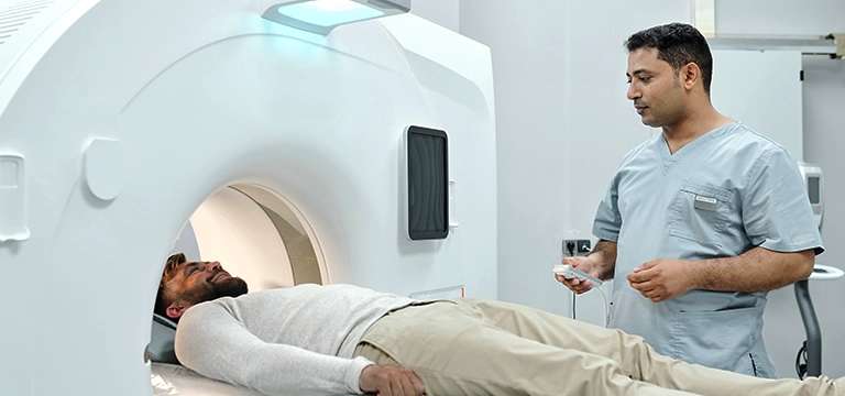
This non-invasive Lumbar Spine MRI scan process starts with the patient lying comfortably on a table that slides into a cylindrical MRI machine. This machine has a strong magnet that creates a magnetic field around the body. When the magnetic field is at work, the protons within the atoms of the body's tissues align themselves in a specific direction. After that, radio waves are then sent into the body, which makes these aligned protons emit signals. These signals are captured by receivers within the MRI machine and processed by a computer to generate detailed images of the lumbar spine.
The conventional and normal Lumbar Spine MRI returns high-resolution images of the anatomy, helping doctors to assess the vertebral bodies, intervertebral discs, spinal cord, nerves, and surrounding soft tissues. However, there are some advanced techniques like functional MRI (fMRI) and diffusion-weighted imaging (DWI), which offer some extra insights into the functional and microstructural aspects of the lumbar spine.
Functional MRI (fMRI): It can measure any changes in blood flow and oxygenation to determine areas of the brain that are activated during specific tasks or stimuli. When it is specifically used for brain imaging, fMRI can also be applied to the lumbar spine to analyze functional connectivity or get responses to certain movements or stimuli.
Diffusion-weighted imaging (DWI): It evaluates the movement of water molecules within tissues. In the lumbar spine, DWI can be of great help in gaining insight into the integrity of nerve fibers and finding out areas of restricted diffusion, which can point toward any nerve compression or damage.
Overall, Lumbar Spine MRI provides a comprehensive and detailed examination of the structures within the lower back. By using these advanced techniques like fMRI and DWI, it facilitates healthcare professionals to visualize anatomical abnormalities and also access functional and microstructural insights. Hence, it leads to more accurate diagnoses and tailored-made treatment approaches for patients.
Different scenarios that call for Lumbar spine MRI
Now we know that Lumbar Spine MRI can be a valuable diagnostic imaging tool carried out in various medical scenarios to investigate and evaluate specific conditions affecting the lower back.
When persistent lower back pain does not improve with conventional treatment, doctors take the help of a lumbar spine MRI to find out the actual causes of it. It can help healthcare professionals to identify the underlying cause of the pain, which may be due to degenerative disc disease, facet joint arthritis, or spinal stenosis.
Another scenario that may warrant a lumbar spine MRI is suspected disc herniation. When doctors suspect a herniated disc, MRI can provide detailed images of the intervertebral discs, allowing them to assess the extent of the herniation, spot the location, and evaluate if it is impinging on nearby nerves. This can be vital information in guiding treatment decisions, whether it is conservative measures or surgical intervention.
On the other hand, lumbar spine MRI is also instrumental in diagnosing spinal cord compression. It can be a boon to understanding ailments like spinal tumors, spinal infections, or spinal cord injuries, which may cause compression of the spinal cord, resulting in neurological deficits.
Doctors can easily view the state of the spinal cord, identify any compression, and assess the extent of the damage with the help of an MRI. This information is very crucial in devising the right management approach, whether it is surgical decompression or other targeted interventions.
Apart from all the above-discussed scenarios, a lumbar spine MRI may also be suggested when healthcare professionals fear spinal fractures or inflammatory conditions like ankylosing spondylitis, or if they want to evaluate the effectiveness of previous spinal surgeries. So, through detailed images of the lumbar spine structures, MRI boosts accurate diagnoses, aids in treatment planning, and facilitates the monitoring of disease progression or treatment outcomes.
Lumbar Spine MRI price
While it is a very important and helpful tool for patients and physicians to enhance treatment outcomes, some people fret about Lumbar Spine MRI's price as well. It is indeed a sophisticated modality and hence does have a price for it.
One must understand that the cost of a Lumbar Spine MRI can vary depending on various factors such as the particular location, the medical facility, and the specific requirements of the patient.
The final Lumbar Spine MRI price may include a host of particulars like the technical fees, professional interpretation, and the use of advanced imaging equipment. On top of it, you must also consider any additional services or contrast agents required during the procedure, which may add more charges.
On the other hand, insurance coverage and healthcare providers may also influence the eventual price. However, despite the varying costs, the value of a Lumbar Spine MRI far outweighs the price. It is invaluable because it provides insights into the complex structures of the lower back, giving accurate diagnoses and helping doctors in the process of formulating effective treatment plans.
Conclusion
To summarize, a Lumbar Spine MRI is touted as an invaluable tool in the field of medical diagnostics, which offers detailed images helping doctors in the analysis of the intricate structures within the lower back. While the price of this modality may vary, its value cannot be overstated. By providing vital information for accurate diagnosis and personalized treatment plans, a Lumbar Spine MRI plays a great role in helping individuals get relief from lumbar spine-related conditions and eventually ultimately boosting their overall well-being and quality of life.
FAQs
Some frequently asked questions on Lumbar Spine and MRI:
What is the role of an MRI in diagnosing lumbar spine conditions?
MRI (Magnetic Resonance Imaging) is a non-invasive imaging modality that helps in diagnosing lumbar spine disorders. It generates detailed images of the structures within the lower back, including the bones, discs, nerves, and soft tissues. Thereby, it helps in identifying abnormalities such as herniated discs, spinal stenosis, tumors, infections, and other conditions affecting the lumbar spine.
How long does it take for a Lumbar Spine MRI?
The duration of a Lumbar Spine MRI is based on several factors such as the facility's protocols and the complexity of the scan. Usually, you can expect the procedure to be completed within 30 minutes to an hour. However, you should also take into account individual cases, which may vary. Some patients may need additional imaging sequences or contrast administration, which could stretch the duration of the scan.
Is the Lumbar Spine MRI procedure painful?
No, the Lumbar Spine MRI procedure itself is not painful. Be assured that it is a non-invasive and painless imaging modality. However, a few individuals may find it discomforting due to lying still for an extended period. If you have any concerns about this potential discomfort, you can talk beforehand with the radiology technologist, who can provide some assistance and ensure your comfort during the scan.


