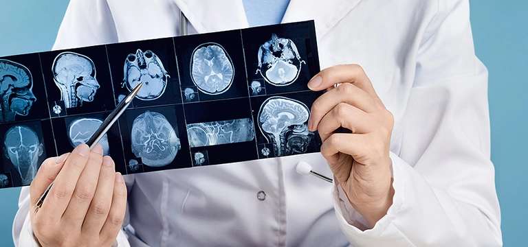
Traumatic Brain Injury (TBI) refers to any injury to the brain that is ensued after an impact from external force It can result from various incidents such as falls, motor vehicle accidents, sports injuries, assaults, or any...
What is Traumatic Brain Injury and what is the Need for Brain MRI in Traumatic Brain Injury?
Traumatic Brain Injury (TBI) refers to any injury to the brain that is ensued after an impact from external force It can result from various incidents such as falls, motor vehicle accidents, sports injuries, assaults, or any other event that causes a significant blow or jolt to the head or body. TBI can range from mild to severe and may have both short-term and long-term effects on the individual.
The impact of a traumatic brain injury can vary widely depending on the severity and location of the injury, as well as the individual's overall health. You might need an MRI brain scan or even an MRI spine scan for this. Some common symptoms and effects of TBI include:
- Physical symptoms: Headaches, dizziness, fatigue, loss of consciousness, seizures, blurred vision, difficulty with balance or coordination, sensory changes, and sleep disturbances.
- Cognitive symptoms: Memory problems, difficulty concentrating or paying attention, slowed thinking, confusion, impaired judgement, and difficulties with problem-solving or decision-making.
- Emotional and behavioral changes: Mood swings, irritability, anxiety, depression, impulsivity, agitation, and changes in social behaviour.
- Communication difficulties: Trouble speaking or understanding language, impaired reading or writing abilities, and challenges with communication in general.
- Sensory changes: Alterations in vision, hearing, taste, or smell, as well as increased sensitivity to light or sound.
In cases of severe TBI, individuals may experience long-lasting or permanent disabilities, such as motor impairments, significant cognitive deficits, or changes in personality and behavior. Recovery from a traumatic brain injury can be a lengthy and complex process, often requiring medical interventions, rehabilitation, and support from healthcare professionals, therapists, and caregivers.
It's important to note that each case of TBI is unique, and the effects can vary greatly from person to person. If you suspect a traumatic brain injury, it is crucial to seek immediate medical attention for an accurate diagnosis and appropriate treatment.
Can there be hidden brain damage in traumatic brain injury?
Yes, it is possible for there to be hidden brain damage in traumatic brain injury (TBI). Hidden brain damage refers to damage that may not be immediately apparent or detectable through routine diagnostic tests, such as CT scans or MRI scans. This type of damage may not be visible on imaging studies but can still have significant effects on the individual's cognitive, emotional, and physical functioning.
Hidden brain damage can occur due to several factors:
- Microscopic damage: TBI can result in microscopic changes to the brain tissue that may not be visible on imaging but can still disrupt the normal functioning of the brain. These microscopic injuries can affect the connections between brain cells, leading to cognitive and behavioral changes.
- Diffuse axonal injury: In some cases of TBI, there may be widespread damage to the axons, which are the long, slender projections that transmit signals between brain cells. Diffuse axonal injury can disrupt communication within the brain and result in cognitive impairments, even if there is no visible structural damage.
- Chemical and metabolic changes: TBI can trigger chemical and metabolic changes in the brain, such as inflammation, neurotransmitter imbalances, and disrupted energy metabolism. These changes can affect brain function without causing obvious structural damage.
- Neuronal death over time: Brain damage following TBI can sometimes progress over time as injured brain cells undergo delayed cell death or degeneration. This ongoing damage may not be immediately evident but can contribute to long-term cognitive decline.
Detecting hidden brain damage can be challenging. Healthcare professionals rely on a combination of clinical assessment, symptom observation, neuropsychological testing, and functional evaluations to identify and evaluate the extent of brain damage in TBI. It is important for individuals with TBI to receive comprehensive evaluations and ongoing monitoring to address both apparent and hidden brain damage and to develop appropriate treatment and rehabilitation plans.
What is the need for head and neck MRI in traumatic brain injury?
Head and neck MRI (Magnetic Resonance Imaging) is often used in cases of traumatic brain injury (TBI) to assess the extent of brain damage and identify any associated injuries or complications. Here are some reasons why a head and neck MRI may be needed:
- Detailed evaluation of brain structures: MRI provides highly detailed images of the brain, allowing healthcare professionals to assess the specific regions affected by the trauma. It can help identify any visible structural damage, such as brain hemorrhages, contusions, or skull fractures. The information obtained from an MRI can assist in determining the severity and location of the brain injury.
- Detection of hidden brain damage: As mentioned earlier, some brain damage may not be immediately apparent and can be missed by other imaging techniques like CT scans. MRI is more sensitive in detecting certain types of brain injuries, such as diffuse axonal injury or small hemorrhages, which might not be visible on a CT scan.
- Evaluation of the neck and spinal cord: Trauma to the head can often be accompanied by injuries to the neck or spinal cord. An MRI of the neck can help identify any damage to the spinal cord, vertebral fractures, or soft tissue injuries. This comprehensive assessment ensures that all potential injuries are considered and appropriate treatment is provided.
- Pre-existing conditions or risk factors: In some cases, individuals with a history of pre-existing conditions or risk factors (such as a brain tumor, vascular malformations, or previous head and neck injuries) may require an MRI to evaluate the baseline status of these structures. This baseline information can help differentiate between pre-existing conditions and new injuries resulting from the traumatic event.
- Post-injury monitoring: MRI can be useful in monitoring the progression of brain injuries over time. It allows healthcare professionals to track changes in the brain, assess healing, and identify any complications or evolving conditions that may require intervention.
It's important to note that the decision to perform a head and neck MRI in cases of traumatic brain injury depends on various factors, including the severity of the injury, clinical presentation, and the judgement of the healthcare provider. The specific imaging approach may vary based on the individual's unique circumstances and the resources available at the medical facility.
Can we get any signs of old TBI in MRI Scan of brain?
In some cases, signs of old traumatic brain injury (TBI) can be detected on a magnetic resonance imaging (MRI) scan. While the acute effects of TBI are often more apparent on imaging shortly after the injury, certain long-term effects or structural changes may still be observable in an MRI conducted months or even years later. However, it's important to note that not all cases of old TBI will exhibit visible signs on an MRI.
Here are some possible signs of old TBI that an MRI might reveal:
- Scarring or gliosis: Following a TBI, the brain may undergo a healing process that involves the formation of scar tissue or an increase in the number of glial cells. These changes can be visible as areas of increased signal intensity on an MRI scan.
- Enlarged ventricles or cortical atrophy: Severe or repeated TBIs can result in brain volume loss, leading to the enlargement of the brain's fluid-filled spaces called ventricles or cortical atrophy. These changes may be detectable on an MRI.
- White matter abnormalities: TBI can cause damage to the white matter tracts in the brain, resulting in disruptions to the structural integrity of these pathways. An MRI can show signs of white matter abnormalities, such as areas of increased signal intensity or abnormal diffusion patterns.
- Microhemorrhages: In some cases, small areas of bleeding (microhemorrhages) may occur in the brain following a TBI. These tiny bleeds can sometimes be seen as small dark spots on certain MRI sequences, such as susceptibility-weighted imaging (SWI).
It's important to note that the visibility of these signs on an MRI can vary depending on various factors, including the severity and location of the initial injury, the time that has elapsed since the injury, and the specific imaging techniques utilized.
It's also worth mentioning that the presence of old TBI-related changes on an MRI should be interpreted in conjunction with the individual's clinical history, symptoms, and other diagnostic findings to form a comprehensive understanding of their condition.
Ultimately, the assessment of old TBI on an MRI is a complex process that requires the expertise of a radiologist or a healthcare professional experienced in interpreting neuroimaging results.
Conclusion
In conclusion, traumatic brain injury (TBI) refers to damage or injury to the brain caused by an external force or trauma. It can range from mild to severe, with a wide range of physical, cognitive, emotional, and behavioral symptoms. While some brain damage may be immediately evident, TBI can also result in hidden or subtle damage that may not be apparent on routine imaging tests. Hidden brain damage can include microscopic injuries, diffuse axonal injury, chemical and metabolic changes, and delayed neuronal death. Head and neck MRI plays a crucial role in the evaluation of TBI by providing detailed images of brain structures, detecting hidden brain damage, assessing the neck and spinal cord, evaluating pre-existing conditions or risk factors, and monitoring the progression of injuries over time. However, not all cases of old TBI will exhibit visible signs on an MRI, and the interpretation of MRI findings should be done in conjunction with the individual's clinical history and other diagnostic assessments. Overall, the detection and understanding of TBI, both apparent and hidden, require comprehensive evaluation and ongoing care from healthcare professionals to provide appropriate treatment and support for individuals affected by this complex condition.


