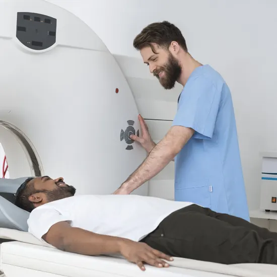
MRI stands for Magnetic Resonance Imaging. It is a medical imaging technique that uses a highly strong magnetic field and also radio waves to obtain the images of the body's internal structures in detail. During an...
What is an MRI Scan?
MRI stands for Magnetic Resonance Imaging. It is a medical imaging technique that uses a highly strong magnetic field and also radio waves to obtain the images of the body's internal structures in detail. During an MRI scan, the patient lies on a table that slides into a tunnel-shaped machine. The machine generates a magnetic field around the body, and radio waves are used to stimulate the body's atoms. The energy released from these atoms is then detected by the machine and used to create images of the body's tissues and organs. MRI scans are non-invasive and do not use ionizing radiation, making them a safe and effective way to diagnose a wide range of conditions, including brain and spinal cord injuries, tumors, and musculoskeletal disorders. Unlike other imaging techniques, such as X-rays and CT scans, MRI scans do not use ionizing radiation, making them a safe and effective diagnostic tool.
What are The Types of MRI Scan?
The type of MRI scan recommended will depend on the specific condition being diagnosed or monitored.
There are several types of MRI scans that are used for different purposes. Some of the most common types of MRI scans include:
- Brain MRI: This type of MRI is used to image the brain and diagnose conditions such as stroke, tumors, and multiple sclerosis.
- Spine MRI: A spine MRI is used to diagnose conditions such as herniated discs, spinal stenosis, and tumors.
- Musculoskeletal MRI: This type of MRI is used to evaluate joints, tendons, ligaments, and muscles for conditions such as arthritis, tears, and sprains.
- Abdominal MRI: An abdominal MRI is used to diagnose conditions such as tumors, liver disease, and gastrointestinal problems.
- Cardiac MRI: This type of MRI is used to evaluate the structure and function of the heart and diagnose conditions such as heart disease and heart attacks.
- Breast MRI: A breast MRI is used to evaluate breast tissue and diagnose conditions such as breast cancer.
- Functional MRI (fMRI): This type of MRI is used to study brain activity and diagnose conditions such as epilepsy and Alzheimer's disease.
What is the Preparation for an MRI scan?
The preparation for an MRI scan can vary depending on the type of scan being performed and the specific instructions from the healthcare provider. However, some general guidelines for MRI preparation include:
- Notify the healthcare provider of any metal in the body, such as pacemakers, implants, or metal fragments, as they may be contraindicated for MRI.
- Avoid wearing clothing with zippers, buttons or snaps. Wear loose, comfortable clothing without metal to the MRI appointment.
- Remove all jewelry, watches, and hair accessories before the scan.
- Let the healthcare provider know if there is a possibility of pregnancy.
- Fast for several hours before the scan if the healthcare provider instructs to do so.
- Take any prescribed medications as usual, unless instructed otherwise by the healthcare provider.
- Inform the healthcare provider of any allergies to contrast dye or medications.
It is important to follow all instructions from the healthcare provider to ensure the MRI scan is safe and accurate.
What are the Indications of MRI Scan?
An MRI (Magnetic Resonance Imaging) scan is a diagnostic tool that uses a powerful magnetic field, radio waves, and a computer to produce detailed images of the body's internal structures.
Here are some common indications for an MRI scan:
- Abdominal pain
- Joint pain or injury
- Headaches
- Neck or back pain
- Spinal cord injury
- Brain or nervous system disorders
- Cancer detection and staging
- Cardiac and vascular abnormalities
- Breast cancer screening
- Prostate cancer screening
- Pelvic pain or abnormal bleeding
- Stroke or TIA (transient ischemic attack)
- Multiple sclerosis
- Arthritis
- Kidney and liver disease
These are just a few examples of the many conditions that may warrant an MRI scan. Your doctor or healthcare provider will determine whether an MRI is necessary based on your individual symptoms, medical history, and physical examination.
What is the time duration of MRI scan?
The duration of an MRI (Magnetic Resonance Imaging) scan can vary depending on the type of scan and the body part being imaged. Generally, an MRI scan can take anywhere from 15 minutes to an hour or more.
For example, a standard MRI of the brain or spine typically takes about 30 to 45 minutes, while an MRI of the abdomen or pelvis can take up to an hour or more. The duration of the scan can also depend on whether or not contrast dye is used, as this may require additional imaging sequences.
It's important to note that during the MRI scan, you will need to lie still on a table while the machine takes images. This can be uncomfortable for some people, especially if you are claustrophobic. If you experience anxiety or discomfort, you can discuss this with your healthcare provider or the imaging center before the scan to see if they can provide any accommodations or sedation to help you feel more comfortable.
What is the Procedure of an MRI Scan?
The procedure for an MRI (Magnetic Resonance Imaging) scan typically involves the following steps:
- Preparation: Before the scan, you will be asked to remove any metal objects or clothing that contain metal, such as jewelry or clothing with zippers, snaps, or buttons. You may be instructed to change clothing into a hospital gown.
- Positioning: You will lie down on a table that slides into the MRI machine. Depending on the body part being scanned, you may be positioned headfirst or feet first. You will be given earplugs or headphones to wear to help block out the noise from the MRI machine.
- Imaging: The MRI machine uses a powerful magnet and radio waves to produce images of the body. You will need to lie still during the scan to prevent any blurring of the images. The technician may also ask you to hold your breath for short periods of time to help improve image quality. The machine may produce loud tapping or knocking noises during the scan, but you can communicate with the technician via an intercom system.
- Contrast dye (if needed): Depending on the reason for the scan, your doctor may request that contrast dye be used to help highlight certain areas of the body. The dye is injected into a vein in your arm before or during the scan.
- Completion: Once the scan is complete, the technician will slide you out of the machine and remove any IV lines or electrodes that were used during the scan. You be able to start again your normal activities immediately after the scan.
After the scan is complete, the images will be reviewed and interpreted by a radiologist, who will send a report to your doctor. Your doctor will then discuss the results with you and determine any necessary follow-up or treatment.
Which medication is to be injected for MRI Scan?
Contrast dye is a type of medication that may be injected during an MRI (Magnetic Resonance Imaging) scan. The contrast dye is a special type of fluid that contains a substance called gadolinium. This substance helps highlight certain areas of the body and makes them easier to see on the MRI images.
The contrast dye is injected into a vein in your arm using a small needle or catheter. You may feel a slight pinch or burning sensation when the needle is inserted, but the injection typically takes only a few seconds.
Not all MRI scans require a contrast dye, and your doctor will determine whether or not it is necessary based on your individual medical needs. If you have a history of kidney disease or allergic reactions to contrast dye, your doctor may choose to use an alternative imaging technique or take precautions to reduce the risk of an adverse reaction.
What are the Price of an MRI Scan?
The cost of an MRI scan can vary depending on several factors such as the type of MRI, the location, and the diagnostic center. In Delhi, the average cost of an MRI without insurance ranges from INR 5000 to INR 50000. However, the cost can be higher for more advanced MRI procedures or if contrast dye is needed.
How much time does it take to Prepare MRI Scan Reports?
The time it takes to prepare an MRI Scan (magnetic resonance imaging) report varies depending on several factors, such as the complexity of the scan, the number of images that need to be reviewed. In general, it can take anywhere from 4 hours to 5 hours for an MRI report to be prepared. Because after an MRI scan is completed, the images are reviewed by the radiologist, who then prepare a report detailing the findings of the scan. The radiologist carefully examines each image and look for any abnormalities or areas of concerns. Once the radiologist has completed their review, they will prepare a written report that includes their findings and any recommendations for further testing or treatment. This report is typically sent to the referring physician, who will discuss the results with the patient.
What is the Role of an MRI Scan in Follow-Up of Patients?
MRI (magnetic resonance imaging) plays an important role in follow-up care for patients who have been diagnosed with various medical conditions, including cancer, neurological disorders, and musculoskeletal injuries. Here are some ways MRI scans are used in follow-up care:
- Assessing treatment response: MRI scans can be used to assess how well a patient is responding to treatment. For example, if a patient has undergone chemotherapy or radiation therapy for cancer, MRI scans can help determine if the tumor is shrinking or if there are any signs of recurrence.
- Detecting progression: MRI scans can help detect disease progression or recurrence in patients who have been previously diagnosed with a medical condition. For example, MRI scans can help detect new tumors or lesions in patients who have a history of cancer.
- Monitoring chronic conditions: MRI scans can be used to monitor chronic conditions such as multiple sclerosis or Alzheimer's disease. Repeat MRI scans can help track the progression of these conditions and guide treatment decisions.
- Identifying complications: MRI scans can help identify complications that may arise from certain medical procedures, such as spinal surgery or joint replacement surgery. MRI scans can help detect infections, fluid build-up, or other complications.
MRI scans are a valuable tool in follow-up care for patients with various medical conditions. MRI scans can help assess treatment response, detect disease progression or recurrence, monitor chronic conditions, and identify complications from medical procedures.
What are the Benefits of MRI Scan?
MRI (magnetic resonance imaging) scan is a non-invasive medical imaging technique that uses a powerful magnetic field, radio waves, and a computer to produce detailed images of internal body structures.
Here are some of the Benefits of MRI Scans:
- Highly detailed images: MRI scans can produce highly detailed images of soft tissues, such as the brain, spinal cord, joints, and organs, which are not always visible with other imaging techniques.
- Non-invasive: MRI scans are a non-invasive procedure that does not involve radiation, making it a safer alternative to other imaging techniques, such as CT scans.
- Diagnosis and monitoring of medical conditions: MRI scans can help diagnose and monitor a variety of medical conditions, such as cancer, strokes, heart and vascular disease, and neurological disorders.
- No exposure to radiation: MRI scan doesn’t use ionizing radiation, which can be harmful in high doses, making it a safer option for patients who require frequent imaging.
What is the Role of MRI in Heart/ Cardiac?
MRI (Magnetic resonance imaging) plays an important role in the evaluation and diagnosis of heart conditions. Here are some ways MRI scans are used in the assessment of heart disease:
- Heart Anatomy: Cardiac MRI provides the detail of the heart and its structure including its chambers, valves as well as blood vessels. These images can help doctors diagnose structural abnormalities such as congenital heart defects or heart valve problems.
- Heart Function: MRI Scan can evaluate the function of the heart including how well it is pumping the blood and how much blood being pumped with each beat. This information can help the doctors diagnose and monitor the cardiomyopathy, heart failure and other heart related conditions that affect heart function.
- Blood Flow: MRI scans can also evaluate blood flow through the heart and blood vessels. This can help diagnose conditions such as coronary artery disease, congenital heart defects, and pulmonary hypertension.
- Viability: MRI scans can also evaluate the viability of heart tissue following a heart attack or other cardiac event. This information can help guide treatment decisions and assess the risk of future cardiac events.
What is the Role of MRI Scan in Cancer Detection?
MRI (magnetic resonance imaging) plays an important role in the detection and diagnosis of cancer. Here are some ways MRI scans are used in cancer detection:
Tumor detection: MRI scans are often used to detect tumors in various parts of the body, including the brain, spine, breast, liver, and prostate. MRI scans can detect small tumors that may not be visible on other imaging techniques.
Staging: MRI scans are used to determine the stage of cancer, which helps doctors determine the appropriate treatment plan. MRI scans can help identify the size and location of the tumor, as well as whether it has spread to nearby tissues or lymph nodes.
Treatment planning: MRI scans can help doctors determine the most effective treatment plan for a patient. For example, MRI scans can help identify if a tumor is operable or if chemotherapy or radiation therapy would be more appropriate.
Monitoring: MRI scans are also used to monitor the progress of cancer treatment. Repeat MRI scans can help determine if the tumor is responding to treatment and if there are any signs of recurrence.


