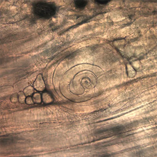
Trichinellosis, often known as trichinosis, is a helminth illness that is mainly spread by eating food that has not been properly prepared. The main causes of infection are pork and its byproducts.
Trichinellosis, often known as trichinosis, is a helminth illness that is mainly spread by eating food that has not been properly prepared. The main causes of infection are pork and its byproducts. Although it often resolves on its own, trichinosis has the potential to be lethal. In nations where pig consumption is high, it is a recurring public health concern.
Trichinosis or trichinellosis is a helminth infection that is typically brought on by subpar or incorrect food preparation. The main causes of infection are pork and its byproducts. Although it usually is a self-limiting disease, it has the potential to be lethal.Sir Richard Owen and Sir James Paget, who saw a mass of worms lining a corpse's diaphragm in 1835, are credited with discovering trichinosis. In nations with high pork consumption, it is a persistent public health concern.
Etiology
A nematode belonging to the genus Trichinella causes trichinosis. Throughout their life cycle inside their host, the parasites shift between skeletal muscle and enteric phases. When the host consumes the infected meat, the enteral phase of the life cycle starts. The larvae are released from the capsule by digestive enzymes in the stomach, where they invade the columnar epithelium of the small intestine. Larvae then move through the tissue, enter the lymphatic system, and are eventually discharged into the bloodstream generally via the thoracic duct. Within 15 days of entering muscle fibres through capillaries, they encyst and turn infectious.
They may stay for the whole of the host's life or for few months. The genus Trichinella contains eleven recognised species. There are two groups among these eleven species: those that encapsulate (are enclosed in a collagen capsule) and infiltrate host muscle cells. The majority of human trichinosis infections and fatalities are caused by Trichinella Spirals, which is a member of the encapsulated group.
Epidemiology
particular, domestic and sylvatic pigs (Sus scrofa) are most commonly affected by Trichinella, the intestinal roundworm that causes trichinosis. Animals that are synanthropic, such as cats, dogs, brown rats, and armadillos, CBC Test are other typical hosts. However, it can also be obtained via wild game, particularly from meat that has not been thoroughly prepared or frozen. Walrus and bear meat have more frequently been linked to trichinosis since 1975.
Trichinella can be found from the tropics all the way up to the Arctic due to its strong infectiousness and the widespread consumption of swine meat. 10,000 trichinosis cases are reported annually, according to the CDC. In the 1940s, there were 400 instances in the United States annually on average; by 2010, there were just 20 cases on average.
Pathophysiology
Three significant changes to cells take place during the acute period of infection:
- 'Nurse cell' production by host cell transformation and removal of sarcomere myofibrils
- If it is an encapsulated species, the larvae become encapsulated.
- A capillary network surrounds the infected cell.
- The cell nucleus is displaced centrally, the sarcoplasm becoming basophilic, and the number and size of the nucleoli increase.
Upon invading inflammatory cells (eosinophils, mast cells, lymphocytes, and monocytes), Trichinella larvae and their metabolites trigger an immune response. The enzymes histaminase and arylsulfatase are released by eosinophils. The production of histamine, serotonin, bradykinin, and prostaglandins PGE2, PGD2, and PGJ2 causes capillaries to become more permeable, which causes tissue edoema, particularly around the eyes.
Vasculitis and microvascular thrombi are other conditions brought on by inflammatory processes. IgE production is increased, which results in allergic reactions such a rash and edoema.
Evaluation
It is simple to misdiagnose the enteral phase since its symptoms are similar to those of other enteral illnesses. In severe infections, larvae are frequently found in muscle biopsy. The worms could be mistaken for pieces of muscle tissue if the biopsy is conducted before the larvae start to coil. When anti-Trichinella IgG antibodies are found, serology reveals a possible trichinosis infection.
Western blot is utilised as a confirming assay from excretory/secretory antigens (ESA), while ELISA is employed as a primary screening test. ESA can identify an illness brought on by any Trichinella taxon, but it cannot identify the precise species.On day 50 after infection, IgG-ELISA reaches 100% sensitivity, but there are many false-negative results in the early stages of the illness.
Symptoms
Trichinosis does not have any pathognomonic symptoms. The present stage of the infection affects the symptoms of Trichinella. The early signs of the enteral (intestinal) phase include nausea, vomiting, upper abdomen pain, low-grade fever, and malaise. These symptoms are brought on by the intestinal invasion of the larvae. After consuming contaminated meat, these start happening 1-2 days later.
Intestinal symptoms go away between two and six weeks after consumption, and parenteral stage symptoms (skeletal muscle) start to show. Periorbital or facial edoema, widespread myalgia, and a state resembling paralysis are only a few of them. Relationship between the number of consumed larvae and illness severity
Treatment
As soon as trichinosis is determined to be the cause of a patient's symptoms, therapy should start. The sickness doesn't progress if it's administered within the first three days of infection. Antihelminthics are the main class of drugs used to treat trichinosis. Albendazole and mebendazole are two of them. In Creatine kinase contrast to mebendazole, whose plasma levels can vary from patient to patient and call for individualised monitoring and dosing, albendazole has been demonstrated to reach appropriate plasma levels and does not require monitoring.
Albendazole should be taken for 8 to 14 days at a dose of 400 mg twice day. Mebendazole should be taken for 3 days at a dosage of 200 to 400 mg three times per day. A second dose of 400 to 500 mg should be administered three times each day for 10 days.
These dosages are suitable for kids and adults, but they should not be used by anyone under the age of two or a pregnant woman. A single dose of 10–20 mg/kg of body weight of Pyrantel can Dehydrogenase provide an option for kids and expectant mothers. It does not harm neonatal or muscle larvae and is solely effective against intestinal larvae. Prednisone, given at a rate of 30-60 mg per day for 10-15 days, is the primary treatment for severe symptoms.
Diagnosis
Eosinophilia and intestinal symptoms are the early signs of trichidiosis infection. Consequently, a number of parasite diseases could be to blame for the symptoms. These consist of:
- Gnathostomiasis
- Strongyloidiasis
- Schistosomiasis
- Hookworm
- Lymphatic filariasis
- Whipworm
Prognosis
The prognosis is dismal for severe instances that involve cardiac or brain problems. Even with treatment, 5% of people with serious infections die. With symptoms going away in 2–6 months in milder cases, the outlook is favourable.
Complications
Complications of acute sickness include vertical infection to the foetus and stillbirths in infected pregnant women. Patients have reported menstruation irregularities, hearing issues, weight loss, hair and nail loss, skin desquamation, aphonia, muscle stiffness, and hoarseness following successful treatment. Creatine Phosphokinase In the first three to five weeks after infection, heart failure or CNS failure can be fatal. Pneumonitis, encephalitis, myocarditis, hypokalemia, blood vessel blockage, and insufficiency of the adrenal glands have all been listed as additional reasons of death.
Generalised myalgia, visual symptoms such conjunctivitis, and other neuropathies are examples of long-term consequences. These could linger for ten years after healing.


