In this article, we will try to explore the significance of pelvic imaging, specifically focusing on the role of MRI in diagnosing and managing pelvic conditions. We will also study what is the purpose of pelvic imaging, and...
Pelvis MRI helps in the diagnosis and management of various conditions affecting the pelvic region. Since there have been remarkable advancements in medical technology, Magnetic Resonance Imaging (MRI) has positioned itself as a powerful tool for churning out detailed assessments of the pelvis.
In this article, we will try to explore the significance of pelvic imaging, specifically focusing on the role of MRI in diagnosing and managing pelvic conditions. We will also study what is the purpose of pelvic imaging, and the specific structures and conditions it can help to analyze. We will try to find out the potential benefits of utilizing MRI for comprehensive pelvic assessment.
So, by understanding the purpose and structure, you will gain valuable insights into the importance of pelvic imaging and how it helps in enhancing diagnostic accuracy and patient outcome.
Preparing for a Pelvis MRI
We should know about the basics of getting ready for a pelvis MRI before going into other details of this imaging technique.
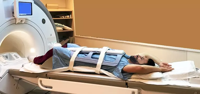
Getting ready for a pelvic MRI starts with a few important steps for ensuring a safe and smooth experience. The healthcare professional will guide the patient through the preparation process, which involves filling out the necessary paperwork, discussing any allergies or implants, and addressing concerns about enclosed spaces.
If you have claustrophobia and feel sensitive to small enclosed spaces, your doctor may prescribe a relaxant to help assuage any stress during the scan. You should also bring a family member or friend with you who can drive you home if a relaxant is administered.
The patient is usually asked not to eat anything before the scan and it will be for a specific period, typically up to four hours. It's essential to empty your bladder two hours before the scan and refrain from using the bathroom until the procedure is completed to avoid potential complications and prolonging the MRI.
One should wear comfortable clothing, such as loungewear or sportswear, and remove any jewelry or valuables at home, in your car, or with your accompanying person. By following these simple steps you can help facilitate a successful pelvic MRI examination.
Understanding the process of Pelvis MRI
Magnetic resonance imaging (MRI) is a highly evolved medical imaging technique that is based on the principle of a strong magnetic field and radio waves to churn detailed and cross-sectional images of the body's internal structures.
Similarly, when it comes to pelvic imaging, MRI has been a highly relevant and invaluable tool for doctors treating patients with pelvis ailments. Owing to its quality to generate high-resolution images, MRI empowers healthcare professionals to gain sufficient access to the pelvis region with exceptional clarity. By providing valuable insights into the organs, bones, blood vessels, and surrounding tissues the pelvis MRI can be a boon for patients.
It is very useful in diagnosing and monitoring disorders such as gynecological problems, pelvic inflammatory disease, uterine fibroids, ovarian cysts, prostate cancer, and rectal tumors, among others. Since this is a non-invasive technique there is the absence of ionizing radiation, MRI is regarded as a safe bet for pelvic imaging. It offers a comprehensive assessment that facilitates accurate diagnosis, treatment planning, and monitoring the effectiveness of treatment interventions.
Conventional MRI vs special type of pelvic MRI scan
There can be some differences between conventional MRI and specialized pelvic MRI techniques. These changes can be significant. While conventional MRI is capable of providing valuable information about the pelvic region, capturing specific images of the organs and structures within, the specialized pelvic MRI modalities can offer enhanced visualization and specific imaging protocols designed to address unique challenges and requirements.
For instance, diffusion-weighted imaging (DWI) and dynamic contrast-enhanced imaging (DCE) are such specialized techniques that provide many additional functional and physiological insights into the tissues.
These techniques boost the assessment of tissue cellularity, perfusion, and vascularity, thereby enhancing the detection and characterization of lesions, particularly in cases of gynecologic malignancies and prostate cancer.
On the other hand, specialized pelvic MRI techniques can make use of multiparametric imaging, which is a combination of different MRI sequences that can provide a more comprehensive evaluation of the pelvic organs. By integrating various sequences, such as T1-weighted, T2-weighted, and contrast-enhanced images, specialized pelvic MRI techniques are powerful diagnostic techniques offering a more accurate picture of pelvic pathology, resulting in improved diagnostic capabilities and optimized patient care.
Latest research and advancements in MRI Pelvis
The onset of newer techniques and advanced imaging sequences and protocols has brought about a profound impact on the field of medical imaging, especially when it comes to pelvic diagnostics. These innovative techniques have upended the way healthcare professionals study and interpret imaging data. Hence, there is improved diagnostic accuracy and level of patient care.
As mentioned earlier, sequences like diffusion-weighted imaging (DWI), dynamic contrast-enhanced imaging (DCE), and magnetic resonance spectroscopy (MRS) have revolutionized MRI of the pelvis. Pelvic pathology has gained so much from these advancements due to their invaluable functional and metabolic information.
DWI helps in the assessment of tissue cellularity and diffusion properties, perfecting the detection and characterization of lesions. On the other hand, DCE provides insights into tissue perfusion and vascularity, which is very crucial for distinguishing benign from malignant lesions and monitoring treatment response.
MRS also comes as a non-invasive technique to gain insights into biochemical changes in tissues, helping in the differentiation of various pathologies.
When combined with improved protocols, these new imaging sequences pave the way for tailored and comprehensive pelvic assessments, resulting in more accurate diagnoses, improved treatment planning, and enhanced patient outcomes.
Every day researchers are working on something to curve new pathways, which will culminate in advanced imaging sequences and protocols. They are relentlessly working, pushing the envelope of pelvic imaging, and empowering healthcare professionals to dispense the highest level of patient care.
MRI Pelvis Price
Are you worried about the cost of an MRI of the pelvis? If MRI Pelvis price is a concern for you, it will help to get some idea about what governs its price. The cost of a pelvis MRI scan depends on a variety of factors, such as the location, the medical facility, and any additional services required.
Another key aspect that can have an impact on the price of a pelvis MRI is insurance coverage. The particular healthcare provider may also influence the final price. Hence, you should get in touch with your insurance provider or healthcare facility for specific information.
If you require additional services such as contrast agents or any specialized imaging sequence, there will be extra charges to incur.
Try to collect some information about any medical facilities that offer discounted rates or payment plans for uninsured or self-pay patients.
Always be aware to ask about any likely hidden costs, such as radiologist interpretation fees or facility fees, so that you are not caught on the wrong foot.
Prices may also differ due to the particular pelvic MRI, such as diagnostic imaging, screening, or follow-up evaluations.
So, try to talk about the cost with your healthcare provider and clarify about any concerns or questions before the MRI scan.
Conclusion
To encapsulate, MRI pelvis is a non-invasive imaging technique that can provide unparalleled cross-sectional images of the pelvic region. It is great for the visualization and evaluation of the organs, bones, blood vessels, and surrounding tissues within the pelvis. Working with the principles of utilizing a strong magnetic field and radio waves, it empowers doctors to assess and diagnose various conditions, such as gynecological disorders, pelvic inflammatory disease, uterine fibroids, ovarian cysts, prostate cancer, and rectal tumors accurately.
It is known for providing high-resolution images and versatility. Hence, it has a crucial role to play in guiding treatment planning, closely following treatment interventions, and improving patient outcomes in the realm of pelvic healthcare.
FAQs
Are there any risks linked to a Pelvis MRI?
Pelvis MRI is touted as a safe and non-invasive technique. There is no exposure to ionizing radiation. However, patients who have certain metallic implants or devices may not be the right candidates to undergo an MRI due to safety reasons.
What is the purpose of a Pelvis MRI?
The purpose of a pelvis MRI is to diagnose and evaluate various conditions affecting the pelvic region. It can be a valuable tool in the assessment of gynecological disorders, pelvic inflammatory disease, uterine fibroids, ovarian cysts, prostate cancer, rectal tumors, and other pelvic abnormalities.
How long does a Pelvis MRI take?
The duration of a Pelvis MRI is governed by factors like the specific protocol and the complexity of the scan. On average, it may take between 30 minutes to an hour to complete the scan.


