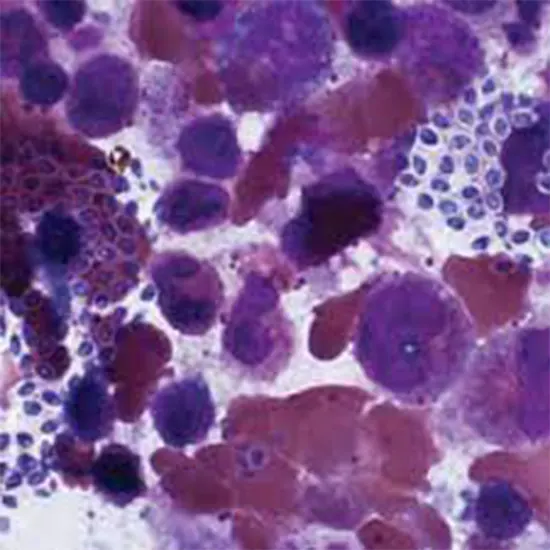
Microsporidia is a bunch of obligated intracellular parasites being the property of the phylum Microsporidia. They are characterized by their unique small size, spore-like structure, and polar tubule that is used to inject the...
Microsporidia is a bunch of obligated intracellular parasites being the property of the phylum Microsporidia. They are characterized by their unique small size, spore-like structure, and polar tubule that is used to inject the infective material into the host cell. They are figured out to contaminate a vast spectrum of hosts, including invertebrates, fish, amphibians, reptiles, birds, and mammals, including humans.
Microsporidia are considered one of the most ancient groups of eukaryotic parasites and have been recognized as a distinct phylum based on molecular and morphological characteristics. They are of significant interest to researchers due to their unique biology and the diseases they cause in humans and animals. In this phylum, over 1400 species of Microsporidia have been identified, and new species are continually being discovered.
Taxonomy and Classification of Microsporidia
Microsporidia is a phylum of unicellular eukaryotic parasites that have been classified based on their molecular and morphological characteristics. The classification of Microsporidia has undergone several revisions in recent years due to advances in molecular phylogenetics.
Currently, Microsporidia are divided into two classes:
Archaeorhizomycetes: This class includes a single species, Archaeorhizomyces finally, which infects marine amoebae.
Microsporidia: This class is further divided into two subclasses, based on differences in their spore formation and morphological features:
Dihaplophasea: This subclass includes species that have a single haploid nucleus within their spores.
Trichomycetes: This subclass includes species that have multiple nuclei within their spores and exhibit a unique coenocytic phase.
The Microsporidia phylum includes over 1400 identified species, which are further classified into 14 orders and over 130 genera. Stool Routine The taxonomic classification of Microsporidia is continually being revised as new species are discovered.
Morphology and Structure of Microsporidia
Microsporidia have a unique morphology and structure that distinguishes them from other groups of eukaryotic parasites.
Morphologically, Microsporidia are characterized by their small size, ranging from 1 to 40 microns in length, and a long polar filament or tubule that is used to inject the infective material into the host cell. The spores of Microsporidia are oval, ellipsoidal, or cylindrical, and are covered Stool Culture by a complex and characteristic wall, consisting of an outer exospore and an inner endospore layer. The exospore layer is thick and resistant, while the endospore layer is thin and delicate.
The structure of Microsporidia is also unique. Microsporidia lack mitochondria and peroxisomes and have a highly modified and reduced cytoplasm. The cytoplasm of Microsporidia is divided into compartments called sporoplasm, which contains the nucleus and other organelles. The nucleus is Gram Stain haploid and contains a small amount of DNA, and ribosomes are attached to the surface of the endoplasmic reticulum. The polar tubule is a structure unique to Microsporidia, and it is used to inject infectious material into the host cell during infection.
In summary, Microsporidia have a unique morphology and structure that distinguishes them from other groups of eukaryotic parasites. Their small size, unique spore wall structure, and lack of mitochondria and peroxisomes, along with the presence of a polar tubule, make them an intriguing group for researchers to study.
Life Cycle of Microsporidia
The life cycle of Microsporidia is complex and involves both asexual and sexual reproduction.
Infection: The life cycle begins when the host ingests a mature Microsporidia spore.
Germination: The spore germinates in the host gut, and the polar tubule is ejected from the spore, injecting the infectious material into the host cell. The infectious material undergoes asexual reproduction in the host cell, producing meronts, which are multinucleated cells that contain many small nuclei called meront nuclei.
Sporogony: The meronts undergo sporogony, a process of asexual reproduction that produces spores containing infectious material.
Release: The mature spores are released from the host cell, and the host cell is destroyed.
Infection: The spores are ingested by a new host, and the life cycle begins again.
In some Microsporidia species, sexual reproduction can also occur, leading to the production of new genetic variations. Sexual reproduction in Microsporidia is known as gametogenesis. The diploid zygote undergoes meiosis, producing haploid spores that can infect new hosts.
The life cycle of Microsporidia can vary between different species, and some species can have a more complex life cycle that involves multiple hosts or life stages. The life cycle of Microsporidia can also vary depending on the x-ray host species that they infect. Overall, Microsporidia have a unique life cycle that involves both asexual and sexual reproduction.
Pathogenesis of Microsporidia
Microsporidia cause disease by damaging host cells and interfering with host metabolism.
The pathogenesis of Microsporidia can be described in three stages:
Invasion: Microsporidia invade host cells using their polar tubule and inject their infectious material. The infectious material can damage host cell membranes and organelles, leading to cell death.
Proliferation: Once inside the host cell, Microsporidia use host cell resources to proliferate and reproduce, leading to further damage to host cells and tissues.
Dissemination: Mature Microsporidia spores are released from infected host cells and can infect new host cells, leading to the spread of infection throughout the host's body.
Diagnosis of Microsporidia
The diagnosis of microsporidiosis can be challenging due to its small size and intracellular location. However, there are several methods available to diagnose microsporidiosis.
Microscopy: The most common method for diagnosing microsporidiosis is microscopy. Staining methods such as Gram, modified trichrome, or calcofluor white can be used to visualize microsporidia spores in clinical specimens. Urine Microscopy is a rapid and inexpensive method, but it may lack specificity in some cases.
Polymerase chain reaction: This can detect the DNA of microsporidia in clinical samples. PCR can catch sight of a low level of infection. PCR-based methods are preferred for the detection of microsporidia in immunocompromised patients.
Serology: Serological tests can detect the presence of antibodies against microsporidia in serum or other body fluids. Serology can help diagnose chronic infections, but it may not be useful for acute infections.
Culture: Microsporidia can be cultured in vitro, but this method is time-consuming and requires special expertise.
In summary, the diagnosis of microsporidia can be made by a combination of microscopy, PCR, and serology. Microscopy is the most common Urine Culture method used, but PCR-based methods are preferred for the detection of microsporidia in immunocompromised patients.
Treatment Options for Microsporidia
Treatment of microsporidiosis (the disease caused by microsporidia) is challenging, as there are limited effective drugs available.
The most commonly used drugs for the treatment of microsporidiosis are albendazole and fumagillin. Albendazole is a broad-spectrum anthelmintic drug that has activity against some microsporidian MRI Brain species. Fumagillin is a potent antibiotic that specifically targets microsporidia. A few more drugs with some success include itraconazole, metronidazole, and azithromycin.
However, drug resistance is a major problem with microsporidia, and treatment failures are common. In addition, some microsporidian infections can cause chronic diseases that are difficult to treat.
Prevention and Control of Microsporidia:
- Good hygiene practices can help prevent microsporidian infections.
- Practice safe sex.
- People with insufficient immunity should take extra precautions to avoid infection.
Significance of Microsporidia in Public Health and Ecology
Microsporidia are significant in both public health and ecology, as they are important parasites that can affect a wide range of organisms.
Here are some specific ways in which microsporidia are significant in public health and ecology:
Public health
Microsporidia can cause disease in humans.
Some microsporidian infections, such as those caused by Enterocytozoon bieneusi, can cause chronic diarrhea, which can be debilitating and life-threatening.
Microsporidia are also important pathogens of animals, including livestock, which can have significant economic impacts.
Ecology
Microsporidia are important parasites of insects and can be used as biological control agents to reduce pest populations.
Some microsporidian species can infect and kill invasive species, making them a potential tool for managing invasive species.
Microsporidia can also affect aquatic organisms, including fish and crustaceans, and can have significant impacts on aquatic ecosystems.
Overall, microsporidia are important organisms that can have significant impacts on both human health and ecological systems. Understanding is significant for formulating useful approaches for their control and management.


