An arthropod-borne flavivirus called the Japanese encephalitis virus (JEV) is common in several Asian, western Pacific, and northern Australian nations. The disease is endemic in 24 countries in Southeast Asia and the...
An arthropod-borne flavivirus called the Japanese encephalitis virus (JEV) is common in several Asian, western Pacific, and northern Australian nations.
The disease is endemic in 24 countries in Southeast Asia and the Western Pacific, putting more than 3 billion people at risk of infection. Symptomatic cases are rare, occurring in about 1 in 250 asymptomatic infections.
Japanese encephalitis (JE) is a major threat with mortality rates of up to 30% in patients with symptomatic disease. Infection causes a variety of clinical illnesses, beginning with flu-like symptoms, neck stiffness, disorientation, coma, seizures, spasticity, and ultimately death.
JEV is one of the greatest public health problems not only because of its large number of deaths, but because of its severe neuropsychiatric sequelae requiring lifelong support that imposes a significant socioeconomic burden.
An outbreak in India (2005) reported 1,700 deaths, most of them children, exacerbating the continuing burden of CBC disease in developing countries. The effectiveness of vector control strategies is limited due to the complex eco-epidemiology of viruses. Vaccination is the most effective preventive measure when JEV is a major public health problem.
This review provides an update on diagnostic methods, preventive and therapeutic management options, and advances in vaccine development.
Transmission
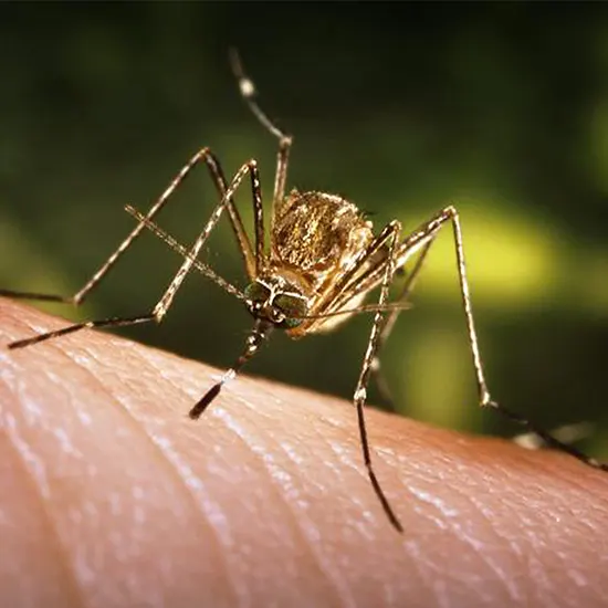
The disease occurs mainly in rural agricultural areas where vector mosquitoes breed near pigs, shorebirds and ducks. The natural cycle of transmission involves several mosquitoes of the genus Culex pipiens, but pigs, birds and bats are susceptible hosts.
The main vector, Culex pipiens, is associated with agricultural practices such as rice cultivation and irrigated crop fields. His increased JEV activity in the new area is attributed to increased population, paddy fields, and pig farming. In EEG addition, aldade birds are thought to be responsible for the long-range spread of JEV and to act as disease reservoirs.
Horses and other non-avian vertebrates are accidental dead hosts because they do not develop sufficient levels of viremia to infect new mosquitoes. Transmission is primarily associated with the rainy season in Southeast Asia, but can occur year-round in tropical regions.
In temperate regions of China, Japan, the Korean Peninsula, and eastern Russia, infections occur mainly in summer and autumn. There are two important determinants of vector density, namely. Precipitation and temperature affect JEV disease burden.
Symptoms
Japanese encephalitis can cause high fever.
Most people with Japanese encephalitis are asymptomatic, but symptoms appear 5 to 15 days after infection.
People with mild Japanese encephalitis may develop only fever and headache, but more serious symptoms can develop rapidly.
Possible symptoms are:
- Headache.
- High fever.
- Shiver.
- Nausea.
- Vomit.
- Torticollis.
- Spasticity.
A person may also experience changes in brain function, such as:
- Stupor.
- Disorientation.
- Coma.
- Convulsions in children.
Pathogen
JEVs are spherical particles of ~50 nm in size and contain an electron-dense core (~30 nm) surrounded by a lipid shell. The genome is a single-stranded positive-sense RNA approximately 11 kb in length.
Viruses consist of a nucleocapsid and a lipoprotein envelope that surrounds the nucleus. The genome is packaged into capsids and encoded by capsid proteins (C). The outer membrane of JEV contains an envelope protein (E) that facilitates entry of the virus into the host cell.
Additionally acknowledged as a protective antigen is the E protein. The genome also encodes a membrane protein (M) and seven nonstructural proteins (NS1, NS2A, NS2B, NS3, NS4A, NS4B, and NS5), the latter of which is a polymerase and the former of which is a helicase.
Antibodies against NS1 are passively transferred to confer protective immunity, but this protein can direct complement-mediated lysis of infected cells through interaction with the cell surface-bound form of NS1.NS2A may be involved in coordinating the shift between RNA packaging and RNA replication. The NS2B CSF protein is a small membrane-bound protein.
Treatment
There is no cure or cure for Japanese encephalitis.
When a person is sick, treatment can only relieve symptoms. Antibiotics are ineffective against viruses and effective antiviral drugs are available.
Prevention is the best treatment for Japanese encephalitis.
Diagnosis
Clinically, it is difficult to distinguish Japanese encephalitis from other encephalitis. In the case of acute encephalitis syndrome (AES), laboratory confirmation is required in such situations.
In Japanese encephalitis, viremia is transient and infection is asymptomatic. Several assays have been developed to detect antibodies induced by natural infection or vaccination. Serological tests include plaque reduction neutralization test (PRNT), hemagglutination inhibition test (HI), indirect immunofluorescence test (IFA), and enzyme-linked immunosorbent test (ELISA).
Various tests based on nucleic acid detection are being investigated for his JEV detection in both human and swine populations. However, laboratory-based surveillance uses the following markers to confirm disease:
- Cerebrospinal fluid (CSF) or serum testing for JEV-specific IgM antibodies is the best procedure for laboratory confirmation.
- Plaque Reduction Neutralization Test.
- Virus isolation.
- Nucleic acid amplification.
Enzyme-linked immunoassay (ELISA)
JEV-specific IgM antibody capture ELISA (MAC-ELISA) is currently the first-line diagnostic assay recommended by WHO for the detection of acute infections. It is a simple platform suitable for rapid testing of large numbers of samples and is considered a cornerstone of clinical practice. The ICMR-National Institute of Virology (NIV) (Pune, India) has developed a reliable IgM capture ELISA kit for rapid diagnosis of her JEV in Sodium serum or CSF samples. Additionally, the kit can detect her IgM for both genotypes. H. GI and GIII, efficient capture.
Reduction of plaque
Neutralization test (PRNT)
It is considered the gold standard for flavivirus diagnostics. PRNT is the test of choice to identify antibodies that may cross-react with other flaviviruses. A 4-fold increase in IgG titers in acute and convalescent sera is considered confirmatory. This increased antibody titer eliminates the possibility of early exposure to the virus.
Virus isolation
Lack of animal models is a long-standing problem in virus isolation. In mice, segregation can be achieved via intracerebral pathways. However, viral isolation is not the optimal method for diagnosis in clinical specimens, as a transient, low-level viremia was observed in her JE infection.
Nucleic acid amplification,RT-PCR tests, quantitative PCR (TaqMan), and restriction fragment length polymorphism (RFLP) analysis are highly specific and sensitive, capable of detecting low copy viruses during the acute or early stages of infection. A useful molecular assay test. The RT-LAMP-LFD assay, which combines reverse transcription loop-mediated isothermal amplification (RT-LAMP) and lateral flow dipstick (LFD), is a rapid, sensitive, and specific assay, making it very useful for diagnosing JEV infection. It is important.
Vaccination
Vaccination is the cheapest means of treatment. Viruses cannot be eliminated because they are maintained in symbiotic cycles with mammals and birds. Vaccination is therefore the most effective way to achieve prevention and long-term protection.
The likelihood of JE control has been revived by improvements in the accessibility and development of JEV vaccinations. The four most common kinds of secure and reliable JE vaccinations are:
- Inactivated Mouse Brain-Derived Vaccines.
- Inactivated Vero Cell Vaccines.
- Live Attenuated Vaccines.
- Chimeric Vaccines.
Antivirals
Approximately 68,000 cases of JE are reported globally each year, despite the fact that only a few licenced vaccines are now available. There are still issues with mortality and morbidity prevention because there are no approved antiviral prophylactic for infection mitigation.
Because of this, safe and affordable antivirals must be developed. Finding effective antivirals for JE required extensive and laborious search efforts. But there aren't any antiviral medications for people that have received FDA approval yet. As a result, supportive treatment is given to people who have Japanese encephalitis.
Several anti-JE drug studies have been conducted. The therapeutic effects of antioxidants such as minocycline, arctigenin, fenofibrate, and curcumin have been previously reported. Clinical studies using dexamethasone, IFN-α2a, and ribavirin did not show encouraging results.
However, the N-methylisatin beta-thiosemicarbazone derivative (SCH 16) has been shown to be effective in inhibiting His JEV in vitro. In another study, minocycline, a semisynthetic tetracycline, reduced the degree of neuronal damage observed with her JEV infection in neuronal cultures by inhibiting oxidative stress.
In addition, minocycline has been shown to maintain blood-brain barrier integrity after JEV infection. A clinical trial of minocycline in Uttar Pradesh, India, found that it was effective in reducing clinical Angiography symptoms such as fever, convulsions, and length of hospital stay. A recent report highlights the utility of bispidine-amino acid conjugates in the treatment of JEV infection due to their unique scaffold properties.
Moreover, in the absence of anti-Japanese encephalitis drugs, combination therapy strategies may be an interesting approach. Effective, safe, and readily available antiviral drugs are a time-consuming need to control mortality in endemic areas around the world.


