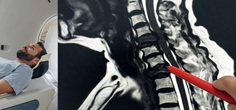
A spine MRI (magnetic resonance imaging) is referred to a non-invasive medical technique that uses powerful magnets and radio waves to generate detailed images of the spine's structures.
Introduction
A spine MRI (magnetic resonance imaging) is referred to a non-invasive medical technique that uses powerful magnets and radio waves to generate detailed images of the spine's structures.
It facilitates the visualization of the vertebrae, discs, spinal cord, nerves, and surrounding soft tissues. The main objective of a spine MRI is to augment the process of diagnosis and evaluation of various spinal conditions and injuries.
It helps doctors and other healthcare professionals with critical information about the spine's anatomy, narrowing the abnormalities such as herniated discs, spinal stenosis, tumors, infections, fractures, and spinal cord injuries.
By securing high-resolution images of the spine, an MRI gives an idea to physicians to accurately identify the source of pain, assess the extent of damage, and devise appropriate treatment strategies. Moreover, a spine MRI is paramount in monitoring the progression of spinal conditions, gauging the success of treatment interventions, and mulling any surgical options when necessary.
A spine MRI can promote patient care, ensure accurate diagnoses, and enable targeted treatment approaches for patients with spinal concerns.
Understanding Spine MRI
To interpret a spine MRI a comprehensive understanding of spinal anatomy is foremost for a layman. The spine, or vertebral column, constitutes individual vertebrae stacked on top of each other, which forms the central support structure of the human body.
The vertebrae are categorized into different regions and they are:
- Cervical (neck)
- Thoracic (mid-back)
- Lumbar (lower back)
- Sacral (pelvic) and
- Coccygeal (tailbone)
Between each vertebra, there are intervertebral discs that act as cushions, providing flexibility and shock absorption. Formed by the vertebral arches, the spinal canal houses and protects the spinal cord and nerve roots.
So, an MRI of the spine provides detailed images of these structures, lending valuable insights into various conditions. With its help, healthcare professionals can identify herniated discs, spinal stenosis, fractures, tumors, infections, and other abnormalities.
MRIs can also shed light on the integrity of the spinal cord, nerve roots, and surrounding soft tissues, supporting in diagnosing spinal cord injuries, nerve impingements, and inflammation. Doctors, equipped with the knowledge of spinal anatomy, can accurately interpret images and make informed diagnoses by correlating MRI findings. Invariably, it leads to appropriate treatment plans and improved patient outcomes.
What are the different types of spine MRI scans?
There are different types of spine MRI scans performed to specifically target and evaluate different regions of the spine, which include the cervical, thoracic, and lumbar regions.
Cervical spine MRI: Its main focus is on the neck region. This scan captures detailed and accurate images of the seven vertebrae that make up the cervical spine, as well as the intervertebral discs, spinal cord, nerve roots, and surrounding soft tissues. It can help in evaluating conditions such as disc herniation, spinal stenosis, cervical spondylosis, and spinal cord compression.
Thoracic spine MRI: It is performed to evaluate the middle portion of the spine, which includes the twelve vertebrae corresponding to the upper back. It can provide great insights into spinal abnormalities, tumors, infections, and spinal cord compression affecting this region.
Lumbar spine MRI: This MRI focuses on the lower back, capturing perfect images of the five vertebrae in the lumbar region, intervertebral discs, spinal cord, nerve roots, and adjacent structures. It is beneficial in diagnosing conditions such as disc degeneration, spinal stenosis, spondylolisthesis, and sciatica.
Healthcare professionals conduct these different types of spine MRI scans and precisely evaluate specific regions of the spine. These MRIs return accurate diagnoses, helping doctors to come up with personalized treatment plans, and effective management of spinal disorders.
Preparing for an MRI of the Spine
Going for an MRI of the spine also involves certain preparations for people so that the process is seamless and devoid of any challenges. For a successful spine MRI imaging, it is important to discuss with your doctor about any existing medical conditions, allergies, or implanted devices, such as pacemakers or metal implants. Remember, divulging these details beforehand determines the safety and suitability of the MRI scan for a patient.
Patients who are about to have a spine MRI are usually asked to wear loose and comfortable clothing, and they may have to change into a hospital gown. They should remove any metal objects, including jewelry, watches, and hair accessories.
These objects may interfere with the MRI's magnetic field. People should also remain on an empty stomach for a specific period before undergoing the scan. It has to be specifically followed when a contrast agent has to be administered.
Doctors usually apprise the patient with specific information depending on individual cases. The patient must follow these instructions very carefully so that the process goes well. With adequate preparation for a spine MRI, patients ensure their safety, succeed in getting improved image quality, and contribute to a successful and accurate diagnosis.
A few common concerns related to Spine MRI
The spine MRI scan also has some common concerns and addressing them is an essential part of the process, which will ensure a comfortable and safe experience.
Patients who have claustrophobia may feel anxious or confined while inside the MRI machine. However, some strategies can alleviate this discomfort.
One such strategy can be an open MRI machine. They have wider openings and increased space, the best alternative for those who struggle with claustrophobia.
Apart, constant communication with the healthcare team is also important. They can provide reassurance and simply explain the process in detail to the patient. They can offer some relaxation techniques or sedation options, if necessary.
One should also discuss metal implants or devices with the healthcare provider in advance. Even though some implants may be MRI-compatible, many of them require further evaluation or alternative imaging methods.
The healthcare team has the best knowledge regarding the safety of the MRI scan and is in a position to provide guidance accordingly. In certain cases where an MRI is complicated, alternative imaging modalities such as CT scans or ultrasound may provide the solution. It helps to openly discuss any concerns or anxieties with the doctors and technicians because they have immense experience in managing these issues over the years. They can come up with tailor-made solutions for the procedure so that patient comfort and safety are maintained.
Spine MRI price
There may be patients who worry about the cost of a spine MRI because it is a sophisticated imaging modality and comes with a price. It is important to understand that spine MRI price is governed by several factors. It depends on the location of the facility, the standard of a particular healthcare facility, and the specific imaging center.
The complexities of the scan along with the number of spinal regions that have to be scanned also have a bearing on the final cost.
Then, there are a few additional considerations like the fees for the radiologist’s interpretation, certain specific facility charges, and any required pre- or post-imaging consultations.
Insurance coverage is also an important element in ascertaining any out-of-pocket expenses for a spine MRI. Usually, insurance plans have varying coverage policies, deductibles, and co-payments that can impact the final cost.
So, it is better to consult the healthcare facility or imaging center directly to discuss the specific pricing for a spine MRI. They can provide detailed cost estimates based on individual circumstances.
Conclusion
So, a spine MRI is an invaluable imaging modality that helps in getting detailed images of the structures within the spine. It is pivotal in diagnosing and evaluating various spinal ailments, guiding treatment decisions, and monitoring how effective the interventions have been. With its high-resolution images, a spine MRI augments in accurately identifying abnormalities, such as disc herniation, spinal stenosis, tumors, and spinal cord injuries. It is a respite for patients because of its non-invasive nature and ability to capture comprehensive views of the spine.
FAQs
How much does a spine MRI typically cost?
The cost of a spine MRI depends on several factors. It is based on the location, particular imaging facility, and the difficulty of the scan. You should talk to the healthcare facility or imaging center directly to get accurate pricing information specifically tailored to your specific situation.
Does my insurance cover the cost of a spine MRI?
Insurance coverage for a spine MRI is also a subjective matter. It depends on your specific insurance plan. Some plans may fully or partially cover the cost of the MRI scan, while others may ask you to co-pay. So, review your particular insurance plan and get in touch contact with your insurance company to learn about any potential out-of-pocket expenses for your spine MRI.


