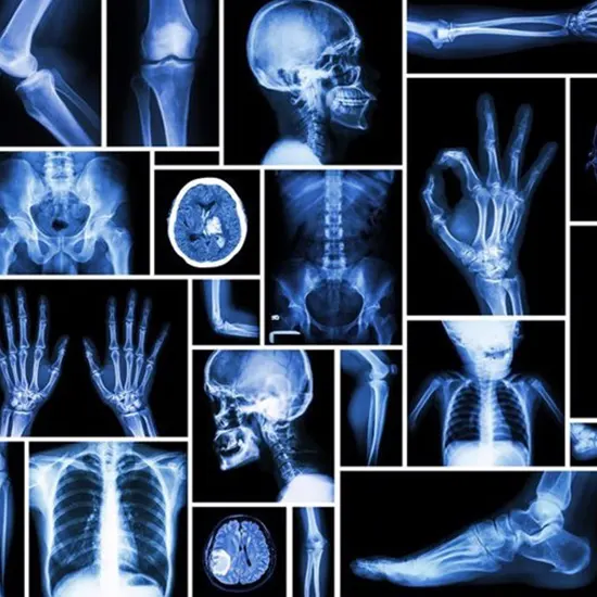
A type of electromagnetic radiation with a high energy that can pass through the body and most other objects. The "shadows" of things inside the body are depicted in an image created by an x-ray that passes through...
A type of electromagnetic radiation with a high energy that can pass through the body and most other objects. The "shadows" of things inside the body are depicted in an image created by an x-ray that passes through the body and strikes an x-ray detector (such as radiographic film or a digital x-ray detector) on the other side of the patient.
A patient is placed in such a way that the area of the body being imaged is situated between an x-ray source and an x-ray detector in order to produce a radiograph. Depending on the radiological density of the tissues they pass through, x-rays that are emitted when the machine is turned on travel through the body and are absorbed in various amounts by various tissues. The atomic number (the quantity of protons in an atom's nucleus) and density of the material being photographed both affect radiological density.
Types of X-Rays
X-ray radiography
Identifies bone fractures, certain tumours and other abnormal masses, pneumonia, some types of traumas, calcifications, foreign objects, or dental issues.
Mammography
Breast radiography is used in mammography to diagnose and detect cancer. On a radiograph, tumours commonly show up as regular or atypically shaped masses that are slightly brighter than the background (i.e., whiter on a black background or blacker on a white background). Micro-calcifications, also known as microscopic pieces of calcium, are detectable with mammograms and appear as extremely bright specks on the image. Even though micro-calcifications are typically benign, certain patterns may be a sign of cancer.
Computed Tomography (CT)
Using traditional x-ray equipment and computer processing, computed tomography (CT) creates a sequence of cross-sectional images of the body that may then be merged to create a three-dimensional x-ray image. The capacity to observe body structures from a variety of angles is provided by CT pictures, which are more detailed than standard radiography.
Fluoroscopy
This technique makes use of x-rays and a fluorescent screen to produce real-time images of bodily activity or to observe diagnostic procedures, such as tracing the course of an injected or swallowed contrast agent. Fluoroscopy, for instance, is used to visualise the activity of a beating heart and, with the use of radiographic contrast chemicals, to visualise blood flow via blood arteries and organs as well as to the heart muscle.
Radiation therapy in cancer
By causing DNA damage to malignant tumours and cells, X-rays and other forms of high-energy radiation can be utilised to treat cancer. Compared to radiation used for diagnostic imaging, radiation utilised for cancer treatment is substantially higher. The source of therapeutic radiation can be an external device, a radioactive substance injected into the bloodstream, or a radioactive substance that is put inside the body, close to or inside tumour cells.
Abdominal X-Ray
An imaging procedure to examine the organs and tissues in the abdomen is an abdominal x-ray. Stomach, intestines, and the spleen are examples of organs.
A KUB (kidneys, ureters, bladder) x-ray is the technical name for the imaging procedure used to examine the bladder and kidney structures.
Barium X-Ray
A particular x-ray of the large intestine, which includes the colon and rectum, is taken during a barium enema.
Extremity X-Ray
The hands, wrists, feet, ankle, leg, thigh, forearm humerus or upper arm, hip, shoulder, or any of these areas can be seen on an extremity x-ray. The word "extremity" is frequently used to describe a human limb.
An image is created on film by an electromagnetic radiation called an X-ray that travels through the body. Bone and other thick structures will appear white. Other structures will be various colours of grey, and the air will be black.
Lumbosacral spine X-Ray
An x-ray of the lumbosacral spine shows the vertebrae, or little bones, that make up the bottom section of the spine. The area that joins the spine to the pelvis, the sacrum, as well as the lumbar region, are included in this region.


