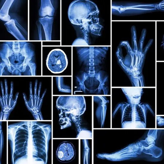
A type of electromagnetic radiation with a high energy that can pass through the body and most other objects. The "shadows" of things inside the body are depicted in an image created by an x-ray that passes through...
Seeing Inside the Chest: An Essential Diagnostic Tool. A chest X-ray is a kind of scientific imaging check that makes use of X-ray science to produce pictures of the chest area. The check produces a black-and-white photograph of the chest that can supply treasured facts about the circumstance of the lungs, heart, bones, and different buildings in the chest.
The Cause of a Chest X-ray
The reason for a chest X-ray is to produce a picture of the chest area through the usage of X-rays. The take look can assist healthcare specialists consider the lungs, heart, bones, and different buildings in the chest. A chest X-ray can be used to diagnose a range of conditions, such as pneumonia, lung cancer, coronary heart disease, and fractures. It can additionally be used to reveal the development of sure prerequisites or to test for any modifications in the chest place over time.
The Step-By-Step Procedure in Performing a Chest X-ray
During a chest X-ray, the affected person stands in front of a unique X-ray machine, whilst a technician positions a flat plate at the back of the patient's back. The technician will ask the affected person to preserve their breath for a few seconds whilst the X-ray is taken. The technique usually takes simply a few minutes and is painless. After the test, the affected person can resume their ordinary activities.
What Sufferers Want To Do To Prepare For a Chest X-ray
In general, there is no exceptional guidance required for a chest X-ray. Patients need to put on loose-fitting apparel and keep away from sporting any earrings or different steel objects that ought to intrude on the imaging process. Patients may additionally be requested to dispose of any apparel or add-ons that cowl the chest area, such as bras or necklaces.
The Viable Dangers and Aspect Results Related To Chest X-rays
Chest X-rays are commonly viewed as secure and do now not have any substantial facet effects. However, like any scientific imaging test, they do expose sufferers to a small quantity of radiation. The quantity of radiation publicity is usually viewed as secure for most patients, however, it might also be a difficulty for pregnant females or sufferers who have gone through more than one imaging exam in the past.
Interpretation of Results
After a chest X-ray, a radiologist will interpret the photos and furnish a record to the patient's healthcare provider. The record may also point out the presence of any abnormalities, such as lung nodules or fluid in the lungs, and furnish suggestions for follow-up trying out or treatment.
Conditions Recognized With Chest X-rays
Chest X-rays can assist diagnose a range of conditions, including:
Pneumonia : An contamination that reasons Pneumonia infection in the lungs.
Lung cancer : A kind of most cancers that starts in the lungs.
Heart disease : A situation that influences the coronary heart and blood vessels.
Fractures : Broken bones in the chest area.
COPD : A crew of lung illnesses that make it hard to breathe.
Alternatives to Chest X-rays
In some cases, healthcare vendors may also advocate different sorts of imaging assessments as a substitute for or in addition to chest X-rays. These might also include:
CT scans : A extra precise kind of imaging check that makes use of X-rays to produce more than one snapshot of the chest.
MRI scans : A non-invasive imaging take a look at that uses a mri test magnetic subject and radio waves to produce pics of the chest.
Ultrasound : A non-invasive imaging Ultrasound check that makes use of sound waves to produce snapshots of the chest.
Follow-up Care
Depending on the effects of a chest X-ray, a healthcare issuer may additionally propose follow-up checking out or treatment. For example, if the X-ray exhibits a viable lung nodule, the company may additionally advocate a CT scan or a biopsy to similarly consider the area. If the X-ray displays fluid in the lungs, the company may additionally propose a cure with a remedy or a system to drain the fluid.
In conclusion, a chest X-ray is a kind of scientific imaging take a look at that makes use of X-ray science to produce photos of the chest area. The take a look at is oftentimes used by way of healthcare companies to consider the circumstance of the lungs, heart, bones, and different constructions in the chest.
A chest X-ray is a painless and non-invasive check that usually takes simply a few minutes to complete. After the test, a radiologist will interpret the pictures and supply a document to the patient's healthcare provider. Depending on the outcomes of the test, the healthcare issuer can also advocate follow-up trying out or treatment.


