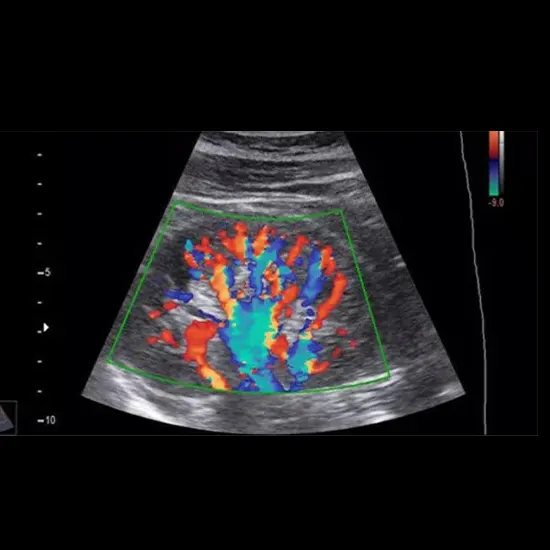
Color Doppler is a medical imaging technique that uses sound waves to visualize blood flow in real-time. It is a non-invasive technique that can provide information about the velocity and direction of blood flow within the...
Color Doppler is a medical imaging technique that uses sound waves to visualize blood flow in real-time. It is a non-invasive technique that can provide information about the velocity and direction of blood flow within the body. For the procedure of color doppler ultrasound, a handheld device(transducer) is placed on the skin over the area of interest. The transducer emits high-frequency sound waves that bounce off the tissues and blood cells within the body. These sound waves are then converted into images using a computer, which can be seen in real-time on a monitor.
In a color Doppler ultrasound, the direction and velocity of blood flow are represented using color-coded images. Blood flowing towards the transducer is shown in shades of red, while blood flowing away from the transducer is shown in shades of blue. The speed of blood flow is represented by the brightness of the color, with brighter colors indicating faster flow.
Color Doppler ultrasound is commonly used to assess blood flow in the heart, blood vessels, and other organs. It can be helpful in diagnosing conditions such as blood clots, narrowing of the arteries, and abnormal blood flow patterns.


