Bone densitometry testing, also known as dual-energy X-ray absorptiometry (DXA or DEXA), is a medical imaging technique used to measure bone mineral density (BMD). It is primarily used to diagnose osteoporosis and assess an...
Bone densitometry testing, or DEXA scanning, is a crucial medical imaging technique used to evaluate bone health and diagnose osteoporosis. With central and peripheral DEXA scans, healthcare providers can assess bone mineral density, identify fracture risk, and guide treatment decisions. Let's explore the types and applications of DEXA scans in detail.
What is Bone Densitometry testing?
Bone densitometry testing, also known as dual-energy X-ray absorptiometry (DXA or DEXA), is a medical imaging technique used to measure bone mineral density (BMD). It is primarily used to diagnose osteoporosis and assess an individual's risk of fractures.
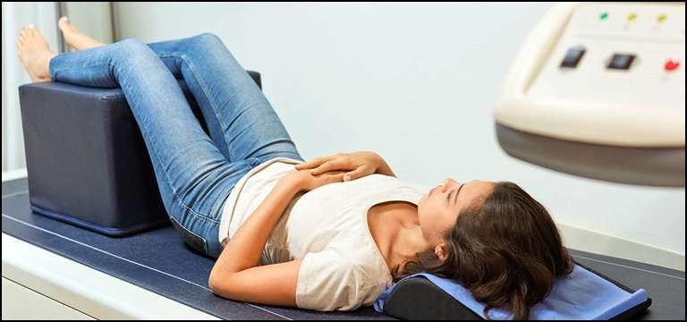
During a bone densitometry test, the patient lies on a table while a specialized X-ray machine scans specific areas of the body, typically the spine, hip, or forearm. The machine emits two low-dose X-ray beams of different energy levels, and the amount of X-ray absorbed by the bones is measured. The test is very quick, non-invasive and is even painless.
Bone densitometry testing is an essential tool for assessing bone health and diagnosing osteoporosis. It helps healthcare providers determine the risk of fractures and develop appropriate treatment plans. The test is often recommended for individuals who are at risk of osteoporosis, such as postmenopausal women, elderly individuals, those with a family history of osteoporosis, and individuals taking certain medications that can affect bone health.
What are the different Types of Dexa Scans?
There are primarily two types of DEXA scans commonly performed:
- Central DEXA Scan: This type of DEXA scan measures bone mineral density (BMD) in the central or axial skeleton, which includes the hip and spine. Central DEXA scans are the most accurate and widely used for diagnosing osteoporosis. They provide valuable information about the risk of fractures in these critical areas.
- Peripheral DEXA Scan: Peripheral DEXA scans measure BMD in peripheral sites, such as the wrist, heel, or finger. These scans are less accurate than central DEXA scans but can still provide valuable information. Peripheral scans are usually used for screening purposes or when central DEXA scans are not feasible or necessary.
Both central and peripheral DEXA scans use dual-energy X-ray absorptiometry technology, but they differ in the areas of the body they evaluate. The choice of the type of DEXA scan depends on the specific clinical situation, the purpose of the test, and the availability of equipment.
It's worth mentioning that there are also specialized variations of DEXA scans that focus on specific areas of interest, such as the trabecular bone score (TBS) or vertebral fracture assessment (VFA). These variations may provide additional information about bone quality or the presence of vertebral fractures, respectively. However, they are not routinely performed as part of standard DEXA scans and may require specialized equipment or software.
When is Axial Dexa Scan Used?
An axial DEXA scan, also known as a central DEXA scan, is used to assess bone mineral density (BMD) in the central or axial skeleton, which includes the hip and spine. This type of DEXA scan is primarily used in the following situations:
- Diagnosis of Osteoporosis: Axial DEXA scans are the most accurate and widely used method for diagnosing osteoporosis. They provide a measurement of BMD in the hip and spine, which are common sites for osteoporotic fractures. The World Health Organization (WHO) defines osteoporosis based on T-scores derived from central DEXA scans.
- Fracture Risk Assessment: Axial DEXA scans can help assess an individual's risk of fractures in the hip and spine. By measuring BMD, healthcare providers can determine the strength and density of bones in these critical areas and evaluate the likelihood of future fractures. The results of the scan, along with other risk factors, help guide treatment decisions.
- Monitoring Response to Treatment: After initiating treatment for osteoporosis, axial DEXA scans may be repeated periodically to monitor the effectiveness of treatment. These follow-up scans can assess changes in BMD over time and determine if the treatment is slowing down bone loss or improving bone density.
- Research and Clinical Trials: Axial DEXA scans are commonly used in research studies and clinical trials focusing on osteoporosis and related bone disorders. These scans provide valuable data for evaluating the efficacy of new treatments, assessing disease progression, and studying bone health in various populations.
It's important to note that axial DEXA scans are more accurate than peripheral DEXA scans for diagnosing osteoporosis and assessing fracture risk in the hip and spine. However, the specific use of axial DEXA scans may vary based on individual patient characteristics, clinical judgment, and local guidelines or practices.
When is Body Dexa Scan Used?
Peripheral DEXA scans, also known as peripheral bone densitometry or pDXA, are used in the following situations:
- Screening: Peripheral DEXA scans are commonly used as a screening tool for assessing bone health in individuals who may be at risk of osteoporosis. They are often performed in community settings or as part of health fairs to identify individuals who may require further evaluation. Screening with peripheral DEXA scans can help identify individuals who may benefit from additional testing or interventions.
- Limited Access to Central DEXA Scans: In some cases, access to central DEXA scanners may be limited, either due to availability or logistical reasons. In such situations, peripheral DEXA scans can be used as an alternative to assess bone mineral density. While peripheral scans are less accurate than central scans, they can still provide useful information, particularly when it is not feasible or necessary to perform a central DEXA scan.
- Monitoring Certain Skeletal Sites: Peripheral DEXA scans are particularly useful for monitoring bone health in specific skeletal sites, such as the wrist, heel, or finger. These scans can help assess BMD changes in these peripheral areas, which may be relevant in certain clinical scenarios or research studies.
- Follow-up and Progression Monitoring: After an initial central DEXA scan, peripheral DEXA scans can be used for follow-up evaluations to monitor changes in BMD over time. This can be helpful in tracking bone health status, evaluating treatment response, or assessing disease progression in specific skeletal regions.
It's important to note that peripheral DEXA scans are generally less accurate than central DEXA scans for diagnosing osteoporosis and assessing fracture risk in the hip and spine. However, they still provide valuable information, especially in screening and monitoring specific peripheral skeletal sites. The choice of using peripheral DEXA scans depends on the clinical context, available resources, and the specific needs of the patient.
How Do I prepare for BMD Dexa Scan?
To prepare for a BMD (bone mineral density) DXA (dual-energy X-ray absorptiometry) scan, you typically don't need to make significant preparations. However, it's important to follow any specific instructions provided by your healthcare provider or the imaging center where the scan will be conducted. Some general guidelines to consider are:
- Clothing: Wear loose, comfortable clothing without metal objects, such as zippers, buttons, or belts, in the area being scanned. Metal can interfere with the scan and affect the accuracy of the results. It is usually recommended to wear clothing without any metal, such as a gown or athletic attire, during the scan.
- Supplements and medications: Inform your healthcare provider about any supplements or medications you are taking, as they may affect the accuracy of the scan or require special precautions. In particular, calcium supplements or medications containing calcium should not be taken on the day of the scan, as they can temporarily affect the results.
- Inform about recent procedures or injections: Let your healthcare provider know if you have recently undergone any contrast agent injections for imaging studies or nuclear medicine scans. These procedures may require a waiting period before a DXA scan can be performed.
- Pregnancy: If you are pregnant or suspect you might be pregnant, inform your healthcare provider. While DXA scans generally involve minimal radiation exposure and are considered safe for adults, they are typically avoided during pregnancy unless the benefits outweigh the potential risks. Alternative imaging methods may be considered in such cases.
- Inform about previous DXA scans: If you have had previous DXA scans done at different facilities, inform your healthcare provider. Having access to previous scan results can help in evaluating changes in bone density over time.
It's important to note that these are general guidelines, and specific instructions may vary depending on the imaging center and your healthcare provider's recommendations.
Conclusion
In conclusion, bone densitometry testing, commonly known as DEXA or DXA scanning, is a valuable medical imaging technique used to assess bone health and diagnose osteoporosis. There are two primary types of DEXA scans: central DEXA scans and peripheral DEXA scans.
Central DEXA scans play a crucial role in diagnosing osteoporosis, assessing fracture risk in critical areas such as the hip and spine, and monitoring treatment effectiveness. They are considered the gold standard for evaluating bone mineral density and are widely used in clinical practice. These scans provide accurate measurements that aid in the identification of individuals at higher risk of fractures and guide appropriate treatment interventions.
On the other hand, peripheral DEXA scans are utilized for screening purposes, monitoring specific peripheral skeletal sites, and when access to central DEXA scans is limited. While they are less accurate than central scans, peripheral DEXA scans still offer valuable information in assessing bone health and identifying individuals who may require further evaluation.
Both central and peripheral DEXA scans contribute to a comprehensive understanding of an individual's bone health status. They help healthcare providers make informed decisions regarding diagnosis, treatment, and monitoring of osteoporosis and fracture risk.


