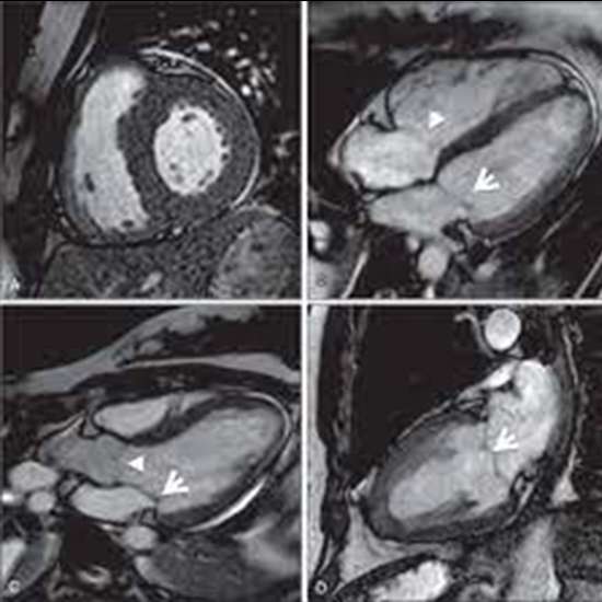
Cardiac MRI is a safe and non-invasive imaging technique that provides detailed information about the structure and function of the heart. It can be used to diagnose a variety of cardiac conditions, such as heart failure,...
Cardiac MRI is a safe and non-invasive imaging technique that provides detailed information about the structure and function of the heart. It can be used to diagnose a variety of cardiac conditions, such as heart failure, heart attacks, and abnormalities of the heart valves and blood vessels.
The procedure for a cardiac MRI (Magnetic Resonance Imaging) involves the following steps:
- Preparation: Before the MRI, you will be asked to remove any metal objects from your body, such as jewelry, watches, and hearing aids. You need to change your dress into a hospital gown for procedure of test. If you have any medical conditions or implants, such as a pacemaker, you should inform your doctor or technologist.
- Positioning: You will be positioned on a sliding table that will move you into the MRI machine. You will need to lie still during the procedure, as movement can affect the quality of the images.
- Electrocardiogram (ECG) gating: A cardiac MRI is usually performed with ECG gating, which means that the MRI is synchronized with your heartbeat. This allows for detailed images of the heart and its structures throughout the cardiac cycle.
- Contrast injection (if necessary): A contrast dye may be injected into a vein in your arm to enhance the images of your heart. The contrast dye can help identify abnormalities such as blockages or damage to the heart muscle.
- Scanning: The MRI machine will make loud banging and thumping noises during the scan. You may be given headphones to block out the noise. The technologist will communicate with you through an intercom system and you will be able to communicate with them as well.
- Completion: The procedure can take between 30 and 90 minutes, depending on the type of scan required. Once the scan is complete, you will be helped off the table.


