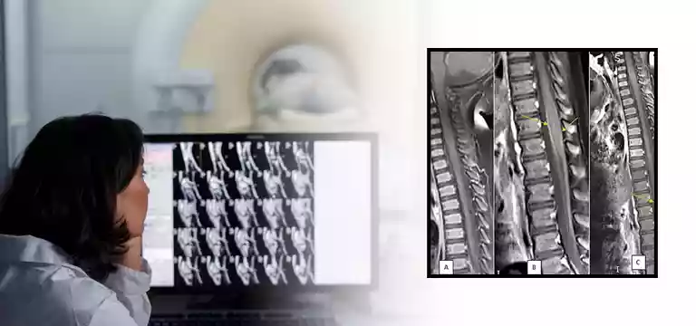
Also known as the thoracic spine, the dorsal spine constitutes twelve vertebrae that connect the cervical spine (neck) and the lumbar spine (lower back). With the help of an MRI (Magnetic Resonance Imaging) of the dorsal spine...
Introduction
Also known as the thoracic spine, the dorsal spine constitutes twelve vertebrae that connect the cervical spine (neck) and the lumbar spine (lower back). With the help of an MRI (Magnetic Resonance Imaging) of the dorsal spine we get valuable diagnostic information on the structures and conditions affecting the middle portion of the vertebral column.
MRI imaging of the dorsal spine offers detailed visualization of the bones, discs, spinal cord, nerve roots, muscles, and surrounding soft tissues. This is a non-invasive and radiation-free imaging technique that comes with superb clarity and precision. It is a great help for healthcare professionals in diagnosing and assessing various dorsal spine pathologies. They get a clear picture of many disorders including herniated discs, degenerative changes, spinal stenosis, tumors, infections, and spinal cord abnormalities.
Through this, we will try to know the significance of MRI in dorsal spine imaging with a special mention of its relevance in diagnosing and guiding appropriate treatment interventions for patients with dorsal spine conditions.
Doctors have been providing optimal care and improved patient outcomes with the help of a dorsal spine MRI.
Let’s understand what a dorsal spine is
The dorsal spine is a vital region of the vertebral column located between the cervical and lumbar spine. As mentioned above it comprises twelve vertebrae, and serves as the protective framework for the vital organs of the chest, such as the heart and lungs. It provides structural stability and allows for limited movement, primarily facilitating rotation and flexion. Moreover, the dorsal spine acts as an attachment site for numerous muscles and ligaments that support posture and facilitate upper body movement.
Disorders of the dorsal spine can range from degenerative conditions, such as osteoarthritis and disc herniation, to congenital abnormalities, fractures, infections, and tumors. Owing to its unique anatomical features and potential implications on a person’s overall health, it is necessary to have a thorough evaluation of the dorsal spine.
In this regard, diagnostic imaging techniques like MRI play an important role in visualizing and assessing the intricate structures within the dorsal spine, facilitating accurate diagnosis, treatment planning, and monitoring of spinal health.
MRI Dorsal Spine: Its Clinical Applications
MRI of the dorsal spine comes with a vast range of clinical applications in diagnosing and managing various conditions affecting this region. One of the primary applications is the evaluation of degenerative changes, such as disc degeneration, facet joint arthritis, and spinal stenosis.
An MRI helps in obtaining a detailed visualization of these structures, helping physicians to know the level of degeneration and how much it has impacted the spinal canal and nerve roots.
Apart from this, MRI is also helpful in diagnosing and characterizing herniated discs, and assessing their size, location, and potential compression of neural structures. It also helps in evaluating any abnormalities in the spinal cord, such as spinal cord compression, myelopathy, and syringomyelia.
It is capable of providing perfect imaging of the spinal cord and surrounding soft tissues, assisting in the examination of any structural abnormalities or masses that may impair neural function. Moreover, MRI is instrumental in detecting and characterizing spinal tumors, both primary and metastatic. It can provide detailed information about the location, size, and extent of the tumor, facilitating treatment planning and monitoring.
For patients who suffer from trauma or fractures, MRI can assess the integrity of the bony structures, ligaments, and soft tissues, helping to guide appropriate management.
Its ability to provide multiplanar imaging and excellent soft tissue contrast is beyond compare, for which, MRI of the dorsal spine continues to be a valuable tool in the diagnosis, treatment, and follow-up of various spinal conditions, invariably improving patient care and outcomes.
New technology and MRI Dorsal Spine
Technology is rapidly developing with new and advanced techniques regularly foraying into the realm of MRI of the dorsal spine. New advancements have revolutionized the imaging and evaluation of spinal pathologies, providing enhanced diagnostic capabilities and improved patient care.
One of the techniques is diffusion-weighted imaging (DWI), which measures the random motion of water molecules within tissues. DWI is an invaluable modality in assessing acute spinal cord injuries, identifying areas of restricted diffusion, and facilitating the characterization of spinal cord pathology.
Apart from this, another advanced technique that is making waves is functional MRI (fMRI), which helps in the assessment of spinal cord function and connectivity. It helps doctors to identify areas of neuronal activation and map neural pathways. It is a great tool to make preoperative planning of spinal cord surgeries.
Meanwhile, magnetic resonance spectroscopy (MRS) is another breakthrough technology that can offer insights into the metabolic changes taking place within the dorsal spine. MRS can assess metabolites such as choline, creatine, and N-acetyl aspartate, contributing to the differentiation between tumor, infection, and inflammation.
Then, there is dynamic contrast-enhanced MRI (DCE-MRI), which sheds valuable light on the vascularity and perfusion of spinal lesions, aiding in the assessment of tumor angiogenesis and treatment response.
It is worth noting that these advanced techniques, along with traditional MRI sequences, allow for a comprehensive evaluation of the dorsal spine. These advanced techniques have always helped in clinical practice by enhancing diagnostic accuracy, improving treatment planning, and optimizing patient outcomes.
It is heartening to observe that the future of MRI in the evaluation of the dorsal spine holds exciting promises. Experts believe that advancements in this area will push the periphery of diagnostic capabilities even further. One fascinating area of development can be witnessed in the realm of quantitative MRI techniques. It provides objective measurements of tissue properties.
Advanced imaging techniques such as diffusion tensor imaging (DTI) and magnetization transfer imaging (MTI) offer the potential to assess microstructural changes and provide insights into the integrity of white matter tracts within the dorsal spine.
There is also a trend of emerging technologies like ultra-high field strength MRI and advanced coil designs. They are capable of achieving higher spatial resolution and improved signal-to-noise ratio, enabling doctors to visualize the dorsal spine with unprecedented clarity and detail.
Furthermore, the use of AI and machine learning may further boost the accuracy and efficiency of image interpretation, helping in the detection and characterization of dorsal spine pathologies. There are also speculations that the future may see the development of novel contrast agents and imaging sequences tailored specifically for the dorsal spine. It will help to detect and monitor subtle abnormalities as well. As there is continuous research, the future of MRI in the evaluation of the dorsal spine looks promising. We can expect enhanced diagnostics, personalized treatments, and improved patient outcomes.
MRI Dorsal Spine price
If you are thinking about the MRI Dorsal Spine price, do some research and investigation so that you get a fair idea about it. It is worth mentioning that the price of an MRI of the Dorsal spine is influenced by a slew of factors. So, what are these factors? One is the location of the facility. Depending on the region where the MRI facility is situated, the price can vary. Another important factor is the complexity involved in the process. At times there is additional sequencing required for a particular MRI. If your scan requires some extra sequencing, expect your MRI Dorsal Spine price to change in this scenario. Talk to your facility, your physician, and others before undergoing the MRI. You will get a clear picture and a fair bit of idea about the pricing before the procedure.
Conclusion
So, we have seen that Dorsal spine MRI is a non-invasive imaging technique that gives detailed images of the middle portion of the vertebral column. Dorsal spine MRI returns high-resolution images of the bones, discs, spinal cord, nerve roots, and surrounding soft tissues. Doctors evaluate a wide range of conditions affecting the dorsal spine, including degenerative changes, herniated discs, spinal stenosis, spinal cord abnormalities, and tumors with the help of a Dorsal Spine MRI.
FAQs
Can a patient undergo a dorsal spine MRI with metallic implants?
While metallic implants may pose challenges during MRI, such as causing artifacts or interfering with the magnetic field, many implants are considered MRI-safe. If a patient has metallic implants he or she should inform the healthcare provider before the MRI. They will be able to guide the compatibility and safety of the procedure.
Is dorsal spine MRI suitable for individuals with claustrophobia?
MRI machines can be confining, causing enough discomfort or anxiety for people with claustrophobia. However, there are techniques to help manage this issue. One should talk to the healthcare provider beforehand in case of such apprehensions about claustrophobia. They will offer strategies such as relaxation techniques or even medication to manage it.


