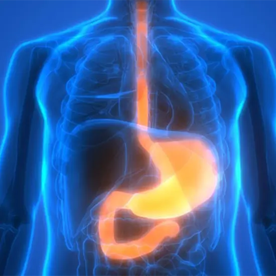
A barium swallow sometimes referred to as an esophagogram, uses a particular type of X-ray to view the upper digestive tract (the esophagus). The test can be done even if you have stomach pain, bloating, vomiting, or difficulty swallowing.
This view is useful in assessing:
• Gastro-esophageal reflux disease (GERD)
• Tracheo-esophageal fistula (TEF done with gastrovedio contrast)
• Dysphasia
• Odynophagia
• Achalasia
• Esophageal hernia
• Esophageal diverticulum
• Aortic mass
• Retrosternal mass/pain
• Esophageal varices
• Esophageal Hold up/ stricture
• Persistent vomiting
• Generalized epigastric pain
• Unexplained weight lose
• Blood in vomiting
• Esophageal ulcer or polyp
The examination method is determined by the study's indication. Overnight fasting from food and the avoidance of smoking and chewing gum to reduce oral and pharyngeal secretions are prerequisites for investigation.
After swallowing high-density barium, the examination is carried out in the upright lateral position. Right lateral views should be obtained first to rule out aspiration or penetration, followed by frontal views. A dynamic video-fluoroscopic examination should be acquired simultaneously for the best evaluation. Spots are quickly obtained during suspended respiration and under phonati.
The preferred method of examination is a double-contrast barium swallow, in which the patient first consumes a packet of effervescent agents before quickly downing a packet of high-density barium. Frontal and left posterior oblique views are then taken, with two exposures focused on the upper/mid oesophagus and two on the distal oesophagus. Next, the table is lowered to the horizontal position, and the patient is turned to the right. The patient is in the prone RAO position and drinking low-density barium.
• Instruct the patient to drink plenty of fluids to prevent constipation.
• Explain to the patient that their face will turn white for two days and not worry about it.