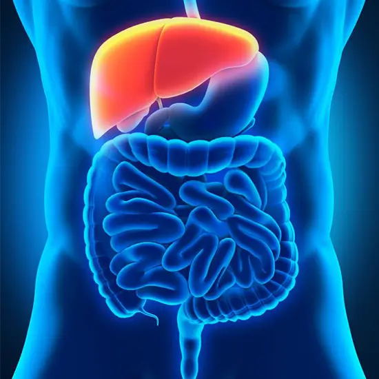
Hepatobiliary imaging in nuclear medicine (HIDA) is an imaging diagnosis based on physiology that uses radioactive medicines and specialised cameras. HIDA radiopharmaceuticals, like bilirubin, are extracted by hepatocytes in the liver and eliminated via the biliary system. The most common indication for HIDA imaging is acute cholecystitis, which is defined by non filling of the gallbladder due to cystic duct obstruction. HIDA can detect high-grade biliary blockage prior to ductal dilatation; images show a prolonged heptagram with no biliary clearance due to high backpressure. HIDA also aids in the diagnosis of partial biliary obstruction caused by stones, biliary stricture, and sphincter of Oddi obstruction. It can demonstrate biliary leakage after cholecystectomy and hepatic transplant.
The gallbladder ejection fraction is calculated after cholecystokinin infusion to identify chronic acalculous gallbladder disease. Gallbladders that are diseased do not contract. There are numerous other, less common but still useful diagnostic applications for HIDA imaging.