Login

X-ray Right Wrist AP & Lateral Views - Test, Procedure & Cost

X-ray Right Wrist AP & Lateral Views

X-ray Right Wrist AP & Lateral Views

Book X-ray Right Wrist AP & Lateral Views Appointment Online Near me at the best price in Delhi/NCR from Ganesh Diagnostic. NABL & NABH Accredited Diagnostic centre and Pathology lab in Delhi offering a wide range of Radiology & Pathology tests. Get Free Ambulance & Free Home Sample collection. 24X7 Hour Open. Call Now at 011-47-444-444 to Book your X-ray Right Wrist AP & Lateral Views at 50% Discount.

₹ 900 ₹ 450

Request A Call Back

What is an X-ray Right Wrist AP & Lateral Views Test?

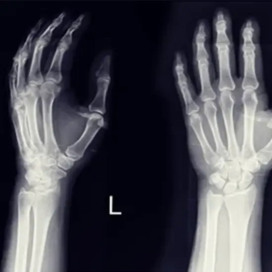
The Right Wrist X-Ray AP and LAT view is an X-ray that would uses small amount of the radiation which will help view the wrist.

It is seen that the wrist comprises of the two bones present in the forearm, namely:

  • Radius
  • Ulna

Eight small bones that make up the- carpal bones.

Why is it done?

This test is generally used to aid in finding infections, the bone cysts, the tumors, and other diseases of your wrist bones.

It would also check the alignment of your bones post surgery or sometimes, even as a part of the bone-age studies that would be before undergoing any surgery.

This lateral wrist radiograph would generally be requested for a myriad of reasons that include the following:

  • Any Trauma
  • Any Suspected Infective Processes
  • Or The Injuries Of The Distal Radius And Ulna
  • Suspected Arthropathy
  • Suspected Foreign Bodies.

The wrist radiographs are considered to be an important as a first-line investigation method in any wrist trauma. This would be a systematic approach to the interpretation being essential.

The failure to identify any fractures of your wrist could result in the range of the complications that include the following:

  • Delayed union
  • Non-union
  • Avascular necrosis
  • Arthritis
  • Joint instability
  • Chronic pain

What is the Procedure for the Right Wrist AP and Lateral View Radiograph Test?

At the hospital site:

  • Change into the hospital gown if requires (otherwise, since this test is only X-ray Right Wrist, you may not be required to change)
  • Remove any jewellery that could hinder X-ray images.
  • During the Test, your body will be positioned in between a machine producing X-rays and a plate. This plate creates the image digitally or using the X-ray film.
  • Depending on the type of X-ray, you may be asked to position your hand in a certain manner. The X-ray technician would then ask you to take a deep breath and hold it for the next few seconds.
  • After the scanning is complete, the respective radiologist would examine the images to understand whether any more of them are required ahead. Once the radiologist is satisfied, the patient could go back home.

Patient Position for PA view and Lateral View

Postero-anterior (PA) view

The PA view would be typically obtained with patient being seated in the following position:

  • The shoulder to be abducted to 90°
  • The elbow flexed to 90° angle
  • The Forearm to be pronated
  • Hand the placed palm down in neutral position with the fingers extended on the designated X-ray plate.

You could also confirm that your hand/wrist is lies in the neutral position by then drawing a line through long axis of your radius, also capitates your  third metacarpal (in the normal axes that are within 10° of line).

Note: Notify the technician in case you are pregnant, or suspect pregnancy

Lateral view

The lateral view of the wrist is technically challenging to interpret due to overlying bony structures.

This view would be obtained by positioning of the patient as follows:

  • The pronated wrist with (ulnar side of fifth digit and facing down onto X-ray plate)
  • The thumb facing upwards.
  • The volar tilt should be between 10°- 25°
  • In this case, this patient position would incorporate your right hand.

After Care

No specific care is required

Cost of Right Wrist AP & Lateral Views Radiology Test in Delhi

At Ganesh Diagnostic and Imaging Centre, we are known for providing excellent service and care to its patients for decades. Lakhs of satisfied patients over the years!

It is an established and renowned diagnostic centre since 2001.

Their excellence is backed by NABH and NABL Accreditations

NABH accreditation is proof of highest standard of care and service provided to the patients. NABL accreditation reflects the competency of laboratories and equipment based on some national and international standards.

The cost of the X-Ray imaging for the Right Wrist AP & LAT Views Test in Delhi starts at INR 450.

Test Type X-ray Right Wrist AP & Lateral Views
Includes

X-ray Right Wrist AP & Lateral Views (X-ray)

Preparation
  • Remove the object that contains Metal, such as Jewellery, Hair Clips, Watches, Rings, or any other similar object from scanning part of the body, so as to avoid a bright or blank spot on the diagnostic film.
  • You must inform the technologist, radiology nurse, and/or physician if you have allergies, are pregnant, breastfeeding, or are on medication.
  • Fasting is not required.
  • Doctor’s Prescription if any
  • Bring all documents related to medical history.
  • Carry Your ID Proof.
Reporting

4-6 hours

Test Price ₹ 450 ₹ 900
Frequently Asked Questions
FAQ

The Radiographic Positioning of Wrist for an X-ray is as follows:

Positioning of the wrist part

  • The Wrist prone is palm up, and you will have to extend your fingers.
  • You should be able to elevate the digits slightly if it is possible to then place your wrist in a close contact to an image receptor.
  • Then, you could have the patient lean in a lateral direction to avoid any wrist rotation.
  • The Central Ray: It will be Perpendicular to an image receptor which is at the center of your wrist.

The wrist series are comprised of the following ways/positions:

  • Poster anterior
  • Oblique
  • Lateral Projection

In one lateral view, the radius and the ulna would be superimposed along with your metacarpals.

The fanning of the fingers and then abducting your thumb would allow for the visualization of the digits with an open joint space.

In an oblique view, your hand would be externally rotated to 45 degrees from a PA position.

A standard wrist procedure for the Wrist X-ray is Posterior-Anterior (PA) and Lateral as follows.

 In any certain circumstances, the further views will be helpful so that 8 overlapping bones can be seen more seen easily.

Wrist is seen to comprise of the following bones:

  • Scaphoid
  • Lunate
  • Triquetrum
  • Pisiform
  • Trapezium
  • Trapezoid
  • Capitate
  • Hamate Bones

A PA view would refer to a film of wrist in which an x-ray beam would passes from your dorsal part to the palmar surface of your wrist, with palmar aspect of wrist lying fiat on an x-ray film.

The AP refers to the wrist positioned with dorsum of your wrist which is touching the film and an x-ray beam that is passing from the palmar to dorsal part.

A 1-2 mm joint space is usually seen between carpals and the metacarpals. In case, the joint space is further narrowed, you could diagnose a carpometacarpal dislocation.

Forearm AP - Position of the patient

  • The patient in this case, must be seated sideways at an end of the table.
  • Then, place your arm on table with your elbow straight.
  • Ideally, your upper arm, elbow, and the forearm would be resting on the table.
  • While, position of the part of supinated hand and your extended elbow.

Median nerve

Your wrist part it is made up of your wrist bones and tough band of the connective tissue (called the ligament).

The median nerve here, would supply your muscles that generally help in bending of the wrist and the fingers.

This is the main nerve for muscles that enable the bending of the thumb.

As such, there aren’t any prescribed limits on the radiation doses for patients.

The doctor generally considers the benefits versus risks for the patient, to understand how much radiation can be justified for accurate diagnosis and monitoring of the disease.

After the patient has undergone any radiographic, or fluoroscopic, or CT, or ultrasound, or the MRI exam, no radiation is said to remain in the body.

At Ganesh Diagnostic and Imaging Centre, we are known for providing excellent service and care to its patients for decades. Lakhs of satisfied patients over the years!

It is an established and renowned diagnostic centre since 2001.

We have 7 centers across Delhi-NCR region; all equipped with the latest and advanced technology.

The rates of scans are reasonably priced. Ganesh Diagnostic and Imaging Centre also offer FLAT 50% OFF on many tests.

Related Tests

Health Packages

Early check ups are always better than delayed ones. Safety, precaution & care is depicted from the several health checkups. Here, we present simple & comprehensive health packages for any kind of testing to ensure the early prescribed treatment to safeguard your health.

view all health packages