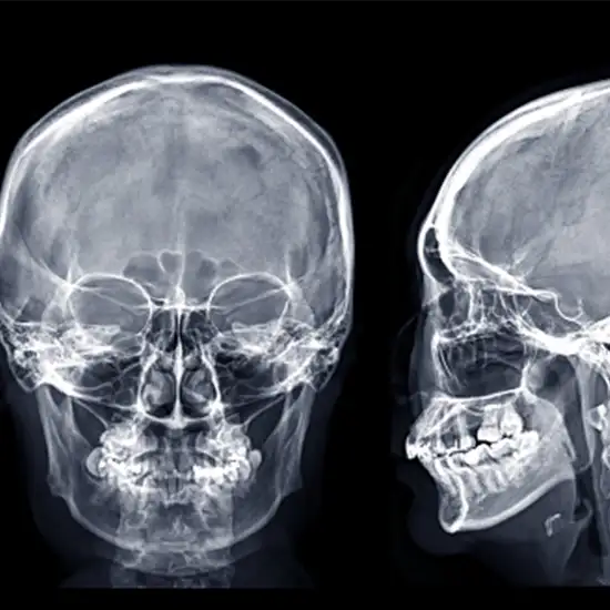
Book X-RAY Skull AP/LAT Appointment Online Near me at the best price in Delhi/NCR from Ganesh Diagnostic. NABL & NABH Accredited Diagnostic centre and Pathology lab in Delhi offering a wide range of Radiology & Pathology tests. Get Free Ambulance & Free Home Sample collection. 24X7 Hour Open. Call Now at 011-47-444-444 to Book your X-RAY Skull AP/LAT at 50% Discount.
The antero posterior AP view/ Lateral View are considered to be specific radiographs of the skull.
This view further offers an overview of the patient’s whole skull rather than striving to just highlight any one region. The “lateral skull base” is known to be one unique area of the skull base; it is situated at the side of the skull.
Antero Posterior View
This Scan helps in the examination for the assessment of the medial and lateral displacements of the skull fractures, in addition to the neoplastic changes and further can lead to Paget’s disease.
Note: As this view would result in a higher radiation dose exposure to the radiosensitive lens of the eyes as compared to the PA view, it is recommended to be used in situations where patient is unable to face the designated detector, like in the trauma settings and for the patients with poor mobility.
This projection is also used to evaluate and look for any skull fractures, in addition to any neoplastic changes and the Paget disease. In trauma settings, a horizontal beam in a lateral projection would demonstrate the air-fluid levels in your sphenoid sinus; this is an indication of the basal skull fracture.
No particular is needed after undergoing the scan
Ganesh Diagnostic and Imaging Centre is a one-stop solution for getting all kinds of tests done, as all services are available under one roof.
The aim of GDIC is to provide world’s finest technology at the lowest price.
The rates of scans are reasonably priced. Ganesh Diagnostic and Imaging Centre also offer FLAT 50% OFF on many tests.
To provide hassle-free diagnostic service we also provide online service to book your appointment. At Ganesh Diagnostic, you can book X-RAY Skull AP/LAT test online by scheduling your appointment. Schedule now for a comprehensive health assessment. We are 24/7 Available at your service.
Patients can rely upon test reports as reports are 100% accurate Cost of X-ray Skull AP LAT Test/Digital in Delhi starts from Rs 350 and upwards, with the variation from one diagnostic center to another.
| Test Type | X-RAY Skull AP/LAT |
| Includes | X-RAY Skull AP/LAT (X-Ray) |
| Preparation |
|
| Reporting | 4-6 hours |
| Test Price |
₹ 350
|

Early check ups are always better than delayed ones. Safety, precaution & care is depicted from the several health checkups. Here, we present simple & comprehensive health packages for any kind of testing to ensure the early prescribed treatment to safeguard your health.