Book Giemsa Stain for Free Living Amoebae Appointment Online Near me at the best price in Delhi/NCR from Ganesh Diagnostic. NABL & NABH Accredited Diagnostic centre and Pathology lab in Delhi offering a wide range of Radiology & Pathology tests. Get Free Ambulance & Free Home Sample collection. 24X7 Hour Open. Call Now at 011-47-444-444 to Book your Giemsa Stain for Free Living Amoebae at 50% Discount.
Lactophenol cotton blue staining for the quick detection of Acanthamoeba cysts in corneal scrapings.] reported using lactophenol cotton blue staining to find Acanthamoeba cysts. This type of stained mount, which is an alternative to Giemsa-stained smears, also enables viewing of amoebae in cultures.
Clinical Diagnosis
Acanthamoeba species: The presence of trophozoites and cysts on stained smears of biopsy specimens (brain tissue, skin, cornea) or corneal scrapings can be used to diagnose an infection with an anthamoeba.
Finding the amoeba in tissue or isolating it provides the conclusive diagnosis of GAE. Both optical and electron microscopy can be used to detect Acanthamoeba trophozoites and cysts in brain tissue, skin lesions, or cerebrospinal fluid (CSF).
Haematoxylin and eosin (H&E), periodic-acid Schiff (PAS), and immunostaining are allegedly the staining methods employed. The effectiveness of these staining techniques in identifying amoebas has not been compared, nevertheless.
Acanthamoeba keratitis must be diagnosed and treated as soon as possible for best results. An eye expert would typically determine the infection's cause based on the patient's symptoms, the ameba's growth after being scraped from the eye, and/or the ability to observe the ameba using confocal microscopy.
Acanthamoeba has a positive or negative Gram score.
Since they are gramme negative and non-acid fast, these organisms cannot be routinely cultivated, yet electron microscopy does show that they are capable of multiplication within the cytoplasm of amoebas.
Giemsa and Wright's stains, both variations of the original Romanowsky stain, are frequently used when examining blood films for parasites.
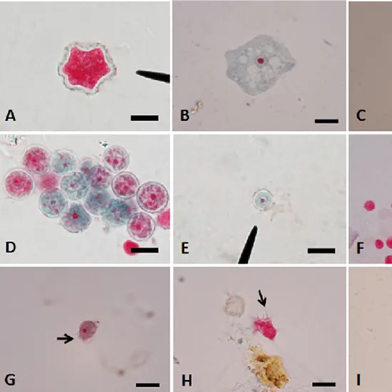
Acanthamoeba cysts were processed for mounting using modified trichrome, Gimenez, and Giemsa staining, and then permanently stained as permanent slides using iodine, eosin, methylene blue, and calcofluor white (CFW) stains.
Chain Reaction of Polymerase (PCR)
To determine whether amebae are present in CSF or tissue, specific molecular instruments can amplify DNA from the amebae.
Protozoa known as free-living amoebae typically reside in the environment and rarely infect human or animal hosts. The two most prevalent species, Acanthamoeba spp. and Naegleria fowleri, both cause disease and infection of the central nervous system.
Amoebas can produce brief cytoplasmic extensions known as pseudopodia, or false feet, which they use as means of locomotion. Amoeboid movement, often known as this sort of movement, is thought to be the first type of animal mobility.
Most of the time, a stool sample may be examined under a microscope to identify this parasite. You might require the antibody test if you have symptoms of amebiasis but the parasite has not been identified in your stool sample or if your doctor suspects that the parasite may have moved outside of your digestive tract.
How is it identified? The most typical method a doctor uses to identify amebiasis is a microscopic examination of feces (poop). The quantity of amoeba being passed in the feces, which changes from day to day, may occasionally be too low to identify from any single sample, necessitating the collection of multiple stool samples.
| Test Type | Giemsa Stain for Free Living Amoebae |
| Includes | Giemsa Stain for Free Living Amoebae (Pathology Test) |
| Preparation | |
| Reporting | Within 24 hours* |
| Test Price |
₹ 300
|

Early check ups are always better than delayed ones. Safety, precaution & care is depicted from the several health checkups. Here, we present simple & comprehensive health packages for any kind of testing to ensure the early prescribed treatment to safeguard your health.