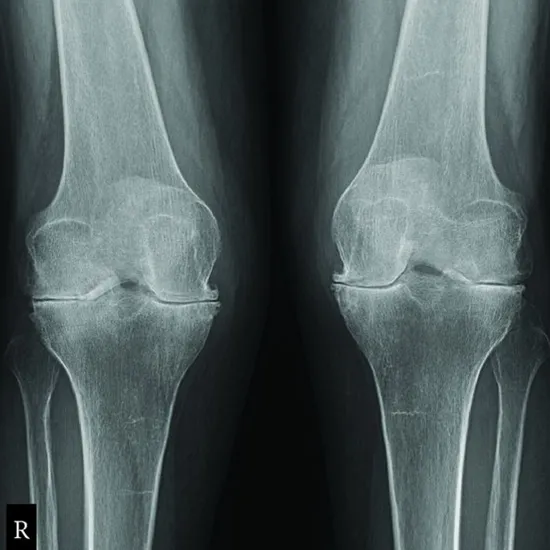
Book X-Ray Right Knee Joint AP Lateral View Appointment Online Near me at the best price in Delhi/NCR from Ganesh Diagnostic. NABL & NABH Accredited Diagnostic centre and Pathology lab in Delhi offering a wide range of Radiology & Pathology tests. Get Free Ambulance & Free Home Sample collection. 24X7 Hour Open. Call Now at 011-47-444-444 to Book your X-Ray Right Knee Joint AP Lateral View at 50% Discount.
The knee antero-posterior and Lateral (AP and LA) projection is considered to be a standard view that is performed to assess a patients’ knee joint and also ones surrounding anatomy.
This is done to get clear image of the patients joint spaces and soft tissues that are around the knee joint.
The anatomical structures that are seen in the x-ray are as follows:
This test is used for the identification of the following:
For being rolled into lateral position, patient would be placed in lateral recumbent position with affected side, towards down.
The affected knee would be flexed to about 20 to 30 degrees. The pelvis must not be rotated at all. The opposite limb would be extended and then placed behind knee which is being examined.
No specific care is required after undergoing the scan
At Ganesh Diagnostic and Imaging Centre, we are known for providing excellent service and care to its patients for decades. Lakhs of satisfied patients over the years!
It is an established and renowned diagnostic centre since 2001.
Their excellence is backed by NABH and NABL Accreditations.
NABH accreditation is proof of highest standard of care and service provided to the patients. NABL accreditation reflects the competency of laboratories and equipment based on some national and international standards.
Test report is available digitally too.
Ganesh Diagnostic and Imaging Centre is a one-stop solution for getting all kinds of tests done, as all services are available under one roof.
The aim of GDIC is to provide world’s finest technology at the lowest price.
The rates of scans are reasonably priced. Ganesh Diagnostic and Imaging Centre also offer FLAT 50% OFF on many tests.
Patients can rely upon test reports as reports are 100% accurate.
The cost of the X-Ray imaging for the Right Knee Joint AP LAT View Test in Delhi starts at INR 450.
Test Type | X-Ray Right Knee Joint AP Lateral View |
Includes | X-ray Right Knee Joint AP Lateral View (X-Ray) |
Preparation |
|
Reporting | 4-6 hours |
Test Price |
₹ 450
|
It is used in combination with the conventional x-rays techniques, Mostly, MRI is considered to be the best choice for examining body's major joints for example, the knee.
The examination is seen to be typically performed to further diagnose or even evaluate the following
Lateral knee view (LA) is considered to be an orthogonal view (angle) of the AP view of the knee.
Projection angle: This projection that requires patient to be able to 'roll' onto one side of his/her knee, thus, it is not appropriate projection in case of trauma, in all the suspected traumatic injuries of knee, horizontal beam of the lateral method could be utilized.
The Technical factors
The right knee should be demonstrated here, and if the x-rays are used for both knees, both knees would have to be demonstrated.
This knee joint space should also be centered in exposure area. The femoral and the tibial Condyles should be symmetrical in nature, indicating no specific rotation.
In the X-ray, the fibular head could also be slightly superimposed on lateral tibial Condyles.
It is seen that a point 2.5 cm below apex of patella is often cited as patient’s center point for the AP of joint, but the patient’s specific measurement will not allow for any variations in the patient build.
The lateral knee-joint X-ray image should meet the requirements as follows:
The position of the patient for rolled lateral position, patient is said to be placed in lateral recumbent position with affected side facing down. The affected knee is also flexed 20 to around 30 degrees.
Keep in mind, the pelvis of the patient should not be rotated. The opposite limb would be extended and also placed behind knee being examined.
Axis of dynamometer was considered to be consistent with axis of motion, and the measurements were made at an angle of 25° and 67° to further examine the differences in either a maximum muscular strength which is according to the joint angle.
Thus, the maximum muscular strength of both the knee-joint extension value, at a value of 67° and flexion value, that is at 25° the value was larger.
The medial side of knee is said to be more congruent and even more stable and medial collateral ligament is also said to be isometric central fibers of the joint. Lateral side of patient’s knee is considered to be more mobile, that slides further in the posterior direction, thus, increasing the flexion and lateral collateral slackens along with the increasing flexion and is thus it is anisometric in nature.
The Four ligaments provide joint stability
The Two collateral (side) ligaments such as the two cruciate ligaments provide knee joint with the support protection from being twisted:
Apart from this. The medial collateral ligament also is known to connect the inner sides of femur and the tibia, which is attached to the joint capsule.
It is known that the 3 most commonly seen injuries are as follows:
Early check ups are always better than delayed ones. Safety, precaution & care is depicted from the several health checkups. Here, we present simple & comprehensive health packages for any kind of testing to ensure the early prescribed treatment to safeguard your health.